P31001
Gene name |
Des |
Protein name |
Desmin |
Names |
|
Species |
Mus musculus (Mouse) |
KEGG Pathway |
mmu:13346 |
EC number |
|
Protein Class |
|
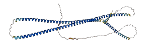
Descriptions
The autoinhibited protein was predicted that may have potential autoinhibitory elements via cis-regPred.
Autoinhibitory domains (AIDs)
Target domain |
|
Relief mechanism |
|
Assay |
cis-regPred |
Accessory elements
No accessory elements
Autoinhibited structure

Activated structure
1 structures for P31001
| Entry ID | Method | Resolution | Chain | Position | Source |
|---|---|---|---|---|---|
| AF-P31001-F1 | Predicted | AlphaFoldDB |
14 variants for P31001
| Variant ID(s) | Position | Change | Description | Diseaes Association | Provenance |
|---|---|---|---|---|---|
| rs3388479309 | 3 | Q>K | No | EVA | |
| rs3388478838 | 21 | A>V | No | EVA | |
| rs13465743 | 30 | L>P | No | EVA | |
| rs3388480420 | 50 | T>R | No | EVA | |
| rs3390181790 | 100 | F>L | No | EVA | |
| rs3388481746 | 103 | T>M | No | EVA | |
| rs36617681 | 154 | Y>* | No | EVA | |
| rs3388481117 | 164 | Q>H | No | EVA | |
| rs3388480789 | 196 | E>D | No | EVA | |
| rs3388479584 | 216 | A>V | No | EVA | |
| rs3388479766 | 316 | A>S | No | EVA | |
| rs3388481734 | 328 | Q>H | No | EVA | |
| rs3388481157 | 418 | P>H | No | EVA | |
| rs3388479657 | 436 | G>D | No | EVA |
No associated diseases with P31001
Functions
14 GO annotations of cellular component
| Name | Definition |
|---|---|
| cardiac myofibril | A cardiac myofibril is a myofibril specific to cardiac muscle cells. |
| cell-cell junction | A cell junction that forms a connection between two or more cells of an organism; excludes direct cytoplasmic intercellular bridges, such as ring canals in insects. |
| contractile fiber | Fibers, composed of actin, myosin, and associated proteins, found in cells of smooth or striated muscle. |
| cytoplasm | The contents of a cell excluding the plasma membrane and nucleus, but including other subcellular structures. |
| cytoskeleton | A cellular structure that forms the internal framework of eukaryotic and prokaryotic cells. The cytoskeleton includes intermediate filaments, microfilaments, microtubules, the microtrabecular lattice, and other structures characterized by a polymeric filamentous nature and long-range order within the cell. The various elements of the cytoskeleton not only serve in the maintenance of cellular shape but also have roles in other cellular functions, including cellular movement, cell division, endocytosis, and movement of organelles. |
| fascia adherens | A cell-cell junction that contains the transmembrane protein N-cadherin, which interacts with identical molecules from neighbouring cells to form a tight mechanical intercellular link; forms a large portion of the intercalated disc, the structure at which myofibrils terminate in cardiomyocytes. |
| gap junction | A cell-cell junction composed of pannexins or innexins and connexins, two different families of channel-forming proteins. |
| intercalated disc | A complex cell-cell junction at which myofibrils terminate in cardiomyocytes; mediates mechanical and electrochemical integration between individual cardiomyocytes. The intercalated disc contains regions of tight mechanical attachment (fasciae adherentes and desmosomes) and electrical coupling (gap junctions) between adjacent cells. |
| intermediate filament cytoskeleton | Cytoskeletal structure made from intermediate filaments, typically organized in the cytosol as an extended system that stretches from the nuclear envelope to the plasma membrane. Some intermediate filaments run parallel to the cell surface, while others traverse the cytosol; together they form an internal framework that helps support the shape and resilience of the cell. |
| neuromuscular junction | The junction between the axon of a motor neuron and a muscle fiber. In response to the arrival of action potentials, the presynaptic button releases molecules of neurotransmitters into the synaptic cleft. These diffuse across the cleft and transmit the signal to the postsynaptic membrane of the muscle fiber, leading to a change in post-synaptic potential. |
| nucleus | A membrane-bounded organelle of eukaryotic cells in which chromosomes are housed and replicated. In most cells, the nucleus contains all of the cell's chromosomes except the organellar chromosomes, and is the site of RNA synthesis and processing. In some species, or in specialized cell types, RNA metabolism or DNA replication may be absent. |
| sarcolemma | The outer membrane of a muscle cell, consisting of the plasma membrane, a covering basement membrane (about 100 nm thick and sometimes common to more than one fiber), and the associated loose network of collagen fibers. |
| type III intermediate filament | A type of intermediate filament, typically made up of one or more of the proteins vimentin, desmin, glial fibrillary acidic protein (GFAP), and peripherin. Unlike the keratins, the type III proteins can form both homo- and heteropolymeric IF filaments. |
| Z disc | Platelike region of a muscle sarcomere to which the plus ends of actin filaments are attached. |
3 GO annotations of molecular function
| Name | Definition |
|---|---|
| cytoskeletal protein binding | Binding to a protein component of a cytoskeleton (actin, microtubule, or intermediate filament cytoskeleton). |
| identical protein binding | Binding to an identical protein or proteins. |
| structural constituent of cytoskeleton | The action of a molecule that contributes to the structural integrity of a cytoskeletal structure. |
3 GO annotations of biological process
| Name | Definition |
|---|---|
| intermediate filament organization | Control of the spatial distribution of intermediate filaments; includes organizing filaments into meshworks, bundles, or other structures, as by cross-linking. |
| muscle organ development | The process whose specific outcome is the progression of the muscle over time, from its formation to the mature structure. The muscle is an organ consisting of a tissue made up of various elongated cells that are specialized to contract and thus to produce movement and mechanical work. |
| skeletal muscle organ development | The progression of a skeletal muscle organ over time from its initial formation to its mature state. A skeletal muscle organ includes the skeletal muscle tissue and its associated connective tissue. |
13 homologous proteins in AiPD
| UniProt AC | Gene Name | Protein Name | Species | Evidence Code |
|---|---|---|---|---|
| P02545 | LMNA | Prelamin-A/C | Homo sapiens (Human) | PR |
| P08670 | VIM | Vimentin | Homo sapiens (Human) | PR |
| P17661 | DES | Desmin | Homo sapiens (Human) | PR |
| P41219 | PRPH | Peripherin | Homo sapiens (Human) | PR |
| P14733 | Lmnb1 | Lamin-B1 | Mus musculus (Mouse) | PR |
| P48678 | Lmna | Prelamin-A/C | Mus musculus (Mouse) | PR |
| P21619 | Lmnb2 | Lamin-B2 | Mus musculus (Mouse) | PR |
| P20152 | Vim | Vimentin | Mus musculus (Mouse) | PR |
| P03995 | Gfap | Glial fibrillary acidic protein | Mus musculus (Mouse) | PR |
| P15331 | Prph | Peripherin | Mus musculus (Mouse) | PR |
| P70615 | Lmnb1 | Lamin-B1 | Rattus norvegicus (Rat) | PR |
| P48679 | Lmna | Prelamin-A/C | Rattus norvegicus (Rat) | PR |
| Q21065 | ifa-3 | Intermediate filament protein ifa-3 | Caenorhabditis elegans | PR |
| 10 | 20 | 30 | 40 | 50 | 60 |
| MSQAYSSSQR | VSSYRRTFGG | APGFSLGSPL | SSPVFPRAGF | GTKGSSSSMT | SRVYQVSRTS |
| 70 | 80 | 90 | 100 | 110 | 120 |
| GGAGGLGSLR | SSRLGTTRAP | SYGAGELLDF | SLADAVNQEF | LATRTNEKVE | LQELNDRFAN |
| 130 | 140 | 150 | 160 | 170 | 180 |
| YIEKVRFLEQ | QNAALAAEVN | RLKGREPTRV | AELYEEEMRE | LRRQVEVLTN | QRARVDVERD |
| 190 | 200 | 210 | 220 | 230 | 240 |
| NLIDDLQRLK | AKLQEEIQLR | EEAENNLAAF | RADVDAATLA | RIDLERRIES | LNEEIAFLKK |
| 250 | 260 | 270 | 280 | 290 | 300 |
| VHEEEIRELQ | AQLQEQQVQV | EMDMSKPDLT | AALRDIRAQY | ETIAAKNISE | AEEWYKSKVS |
| 310 | 320 | 330 | 340 | 350 | 360 |
| DLTQAANKNN | DALRQAKQEM | MEYRHQIQSY | TCEIDALKGT | NDSLMRQMRE | LEDRFASEAN |
| 370 | 380 | 390 | 400 | 410 | 420 |
| GYQDNIARLE | EEIRHLKDEM | ARHLREYQDL | LNVKMALDVE | IATYRKLLEG | EESRINLPIQ |
| 430 | 440 | 450 | 460 | ||
| TFSALNFRET | SPEQRGSEVH | TKKTVMIKTI | ETRDGEVVSE | ATQQQHEVL |