Q9Z0V2
Gene name |
Kcnd2 (Kiaa1044, MNCb-7013) |
Protein name |
Potassium voltage-gated channel subfamily D member 2 |
Names |
Voltage-gated potassium channel subunit Kv4.2 |
Species |
Mus musculus (Mouse) |
KEGG Pathway |
mmu:16508 |
EC number |
|
Protein Class |
|
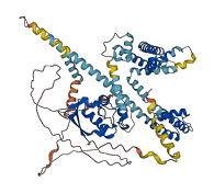
Descriptions
The autoinhibited protein was predicted that may have potential autoinhibitory elements via cis-regPred.
Autoinhibitory domains (AIDs)
Target domain |
|
Relief mechanism |
|
Assay |
cis-regPred |
Accessory elements
No accessory elements
Autoinhibited structure

Activated structure
1 structures for Q9Z0V2
| Entry ID | Method | Resolution | Chain | Position | Source |
|---|---|---|---|---|---|
| AF-Q9Z0V2-F1 | Predicted | AlphaFoldDB |
19 variants for Q9Z0V2
| Variant ID(s) | Position | Change | Description | Diseaes Association | Provenance |
|---|---|---|---|---|---|
| rs3388806338 | 8 | W>S | No | EVA | |
| rs3395793377 | 74 | F>S | No | EVA | |
| rs3388811636 | 74 | F>V | No | EVA | |
| rs3396457113 | 75 | F>L | No | EVA | |
| rs3388812208 | 81 | Q>H | No | EVA | |
| rs3388812192 | 148 | L>I | No | EVA | |
| rs3388819644 | 179 | H>N | No | EVA | |
| rs3412971986 | 204 | V>M | No | EVA | |
| rs3388819630 | 235 | A>V | No | EVA | |
| rs3388812261 | 387 | G>V | No | EVA | |
| rs3388785418 | 450 | N>T | No | EVA | |
| rs3388817315 | 456 | Q>H | No | EVA | |
| rs3388818267 | 480 | H>L | No | EVA | |
| rs3388797255 | 510 | N>K | No | EVA | |
| rs3388823418 | 542 | N>K | No | EVA | |
| rs3388785412 | 551 | G>A | No | EVA | |
| rs3388818204 | 588 | C>S | No | EVA | |
| rs3388817325 | 610 | G>A | No | EVA | |
| rs3396597057 | 631 | L>= | No | EVA |
No associated diseases with Q9Z0V2
5 regional properties for Q9Z0V2
| Type | Name | Position | InterPro Accession |
|---|---|---|---|
| domain | BTB/POZ domain | 41 - 140 | IPR000210 |
| domain | Potassium channel tetramerisation-type BTB domain | 43 - 132 | IPR003131 |
| domain | Ion transport domain | 186 - 416 | IPR005821 |
| domain | Shal-type voltage-gated potassium channels, N-terminal | 3 - 30 | IPR021645 |
| domain | Potassium channel, voltage dependent, Kv4, C-terminal | 472 - 545 | IPR024587 |
Functions
25 GO annotations of cellular component
| Name | Definition |
|---|---|
| anchoring junction | A cell junction that mechanically attaches a cell (and its cytoskeleton) to neighboring cells or to the extracellular matrix. |
| caveola | A membrane raft that forms small pit, depression, or invagination that communicates with the outside of a cell and extends inward, indenting the cytoplasm and the cell membrane. Examples include flask-shaped invaginations of the plasma membrane in adipocytes associated with caveolin proteins, and minute pits or incuppings of the cell membrane formed during pinocytosis. Caveolae may be pinched off to form free vesicles within the cytoplasm. |
| dendrite | A neuron projection that has a short, tapering, morphology. Dendrites receive and integrate signals from other neurons or from sensory stimuli, and conduct nerve impulses towards the axon or the cell body. In most neurons, the impulse is conveyed from dendrites to axon via the cell body, but in some types of unipolar neuron, the impulse does not travel via the cell body. |
| dendritic spine | A small, membranous protrusion from a dendrite that forms a postsynaptic compartment, typically receiving input from a single presynapse. They function as partially isolated biochemical and an electrical compartments. Spine morphology is variable:they can be thin, stubby, mushroom, or branched, with a continuum of intermediate morphologies. They typically terminate in a bulb shape, linked to the dendritic shaft by a restriction. Spine remodeling is though to be involved in synaptic plasticity. |
| GABA-ergic synapse | A synapse that uses GABA as a neurotransmitter. These synapses are typically inhibitory. |
| glutamatergic synapse | A synapse that uses glutamate as a neurotransmitter. |
| integral component of membrane | The component of a membrane consisting of the gene products and protein complexes having at least some part of their peptide sequence embedded in the hydrophobic region of the membrane. |
| integral component of plasma membrane | The component of the plasma membrane consisting of the gene products and protein complexes having at least some part of their peptide sequence embedded in the hydrophobic region of the membrane. |
| integral component of postsynaptic membrane | The component of the postsynaptic membrane consisting of the gene products and protein complexes having at least some part of their peptide sequence embedded in the hydrophobic region of the membrane. |
| integral component of postsynaptic specialization membrane | The component of the postsynaptic specialization membrane consisting of the gene products and protein complexes having at least some part of their peptide sequence embedded in the hydrophobic region of the membrane. |
| intrinsic component of plasma membrane | The component of the plasma membrane consisting of the gene products and protein complexes having either part of their peptide sequence embedded in the hydrophobic region of the membrane or some other covalently attached group such as a GPI anchor that is similarly embedded in the membrane. |
| membrane | A lipid bilayer along with all the proteins and protein complexes embedded in it an attached to it. |
| neuron projection | A prolongation or process extending from a nerve cell, e.g. an axon or dendrite. |
| neuronal cell body | The portion of a neuron that includes the nucleus, but excludes cell projections such as axons and dendrites. |
| neuronal cell body membrane | The plasma membrane of a neuron cell body - excludes the plasma membrane of cell projections such as axons and dendrites. |
| perikaryon | The portion of the cell soma (neuronal cell body) that excludes the nucleus. |
| perinuclear endoplasmic reticulum | The portion of endoplasmic reticulum, the intracellular network of tubules and cisternae, that occurs near the nucleus. The lumen of the perinuclear endoplasmic reticulum is contiguous with the nuclear envelope lumen (also called perinuclear space), the region between the inner and outer nuclear membranes. |
| plasma membrane | The membrane surrounding a cell that separates the cell from its external environment. It consists of a phospholipid bilayer and associated proteins. |
| plasma membrane raft | A membrane raft that is part of the plasma membrane. |
| postsynaptic density | An electron dense network of proteins within and adjacent to the postsynaptic membrane of an asymmetric, neuron-neuron synapse. Its major components include neurotransmitter receptors and the proteins that spatially and functionally organize them such as anchoring and scaffolding molecules, signaling enzymes and cytoskeletal components. |
| postsynaptic membrane | A specialized area of membrane facing the presynaptic membrane on the tip of the nerve ending and separated from it by a minute cleft (the synaptic cleft). Neurotransmitters cross the synaptic cleft and transmit the signal to the postsynaptic membrane. |
| potassium channel complex | An ion channel complex through which potassium ions pass. |
| sarcolemma | The outer membrane of a muscle cell, consisting of the plasma membrane, a covering basement membrane (about 100 nm thick and sometimes common to more than one fiber), and the associated loose network of collagen fibers. |
| T-tubule | Invagination of the plasma membrane of a muscle cell that extends inward from the cell surface around each myofibril. The ends of T-tubules make contact with the sarcoplasmic reticulum membrane. |
| voltage-gated potassium channel complex | A protein complex that forms a transmembrane channel through which potassium ions may cross a cell membrane in response to changes in membrane potential. |
7 GO annotations of molecular function
| Name | Definition |
|---|---|
| A-type (transient outward) potassium channel activity | Enables the transmembrane transfer of a potassium ion by an outwardly-rectifying voltage-gated channel that produces a transient outward current upon a step change in membrane potential. |
| ion channel activity | Enables the facilitated diffusion of an ion (by an energy-independent process) by passage through a transmembrane aqueous pore or channel without evidence for a carrier-mediated mechanism. May be either selective (it enables passage of a specific ion only) or non-selective (it enables passage of two or more ions of same charge but different size). |
| metal ion binding | Binding to a metal ion. |
| potassium channel activity | Enables the facilitated diffusion of a potassium ion (by an energy-independent process) involving passage through a transmembrane aqueous pore or channel without evidence for a carrier-mediated mechanism. |
| protein-containing complex binding | Binding to a macromolecular complex. |
| voltage-gated ion channel activity involved in regulation of postsynaptic membrane potential | Any voltage-gated ion channel activity that is involved in regulation of postsynaptic membrane potential. |
| voltage-gated potassium channel activity | Enables the transmembrane transfer of a potassium ion by a voltage-gated channel. A voltage-gated channel is a channel whose open state is dependent on the voltage across the membrane in which it is embedded. |
9 GO annotations of biological process
| Name | Definition |
|---|---|
| action potential | A process in which membrane potential cycles through a depolarizing spike, triggered in response to depolarization above some threshold, followed by repolarization. This cycle is driven by the flow of ions through various voltage gated channels with different thresholds and ion specificities. |
| cardiac muscle cell action potential | An action potential that occurs in a cardiac muscle cell. |
| cellular response to hypoxia | Any process that results in a change in state or activity of a cell (in terms of movement, secretion, enzyme production, gene expression, etc.) as a result of a stimulus indicating lowered oxygen tension. Hypoxia, defined as a decline in O2 levels below normoxic levels of 20.8 - 20.95%, results in metabolic adaptation at both the cellular and organismal level. |
| locomotor rhythm | The rhythm of the locomotor activity of an organism during its 24 hour activity cycle. |
| neuronal action potential | An action potential that occurs in a neuron. |
| potassium ion transmembrane transport | A process in which a potassium ion is transported from one side of a membrane to the other. |
| protein homooligomerization | The process of creating protein oligomers, compounds composed of a small number, usually between three and ten, of identical component monomers. Oligomers may be formed by the polymerization of a number of monomers or the depolymerization of a large protein polymer. |
| regulation of ion transmembrane transport | Any process that modulates the frequency, rate or extent of the directed movement of ions from one side of a membrane to the other. |
| sensory perception of pain | The series of events required for an organism to receive a painful stimulus, convert it to a molecular signal, and recognize and characterize the signal. Pain is medically defined as the physical sensation of discomfort or distress caused by injury or illness, so can hence be described as a harmful stimulus which signals current (or impending) tissue damage. Pain may come from extremes of temperature, mechanical damage, electricity or from noxious chemical substances. This is a neurological process. |
12 homologous proteins in AiPD
| UniProt AC | Gene Name | Protein Name | Species | Evidence Code |
|---|---|---|---|---|
| Q61423 | Kcna4 | Potassium voltage-gated channel subfamily A member 4 | Mus musculus (Mouse) | PR |
| P16390 | Kcna3 | Potassium voltage-gated channel subfamily A member 3 | Mus musculus (Mouse) | PR |
| P16388 | Kcna1 | Potassium voltage-gated channel subfamily A member 1 | Mus musculus (Mouse) | PR |
| Q80XM3 | Kcng4 | Potassium voltage-gated channel subfamily G member 4 | Mus musculus (Mouse) | PR |
| Q03717 | Kcnb1 | Potassium voltage-gated channel subfamily B member 1 | Mus musculus (Mouse) | PR |
| Q8K3F6 | Kcnq3 | Potassium voltage-gated channel subfamily KQT member 3 | Mus musculus (Mouse) | PR |
| O35174 | Kcns2 | Potassium voltage-gated channel subfamily S member 2 | Mus musculus (Mouse) | PR |
| A6H8H5 | Kcnb2 | Potassium voltage-gated channel subfamily B member 2 | Mus musculus (Mouse) | PR |
| P97414 | Kcnq1 | Potassium voltage-gated channel subfamily KQT member 1 | Mus musculus (Mouse) | PR |
| Q17ST2 | Kcna7 | Potassium voltage-gated channel subfamily A member 7 | Mus musculus (Mouse) | PR |
| Q9Z0V1 | Kcnd3 | Potassium voltage-gated channel subfamily D member 3 | Mus musculus (Mouse) | PR |
| Q7TSH7 | Kcnf1 | Potassium voltage-gated channel subfamily F member 1 | Mus musculus (Mouse) | PR |
| 10 | 20 | 30 | 40 | 50 | 60 |
| MAAGVAAWLP | FARAAAIGWM | PVASGPMPAP | PRQERKRTQD | ALIVLNVSGT | RFQTWQDTLE |
| 70 | 80 | 90 | 100 | 110 | 120 |
| RYPDTLLGSS | ERDFFYHPET | QQYFFDRDPD | IFRHILNFYR | TGKLHYPRHE | CISAYDEELA |
| 130 | 140 | 150 | 160 | 170 | 180 |
| FFGLIPEIIG | DCCYEEYKDR | RRENAERLQD | DADTDNTGES | ALPTMTARQR | VWRAFENPHT |
| 190 | 200 | 210 | 220 | 230 | 240 |
| STMALVFYYV | TGFFIAVSVI | ANVVETVPCG | SSPGHIKELP | CGERYAVAFF | CLDTACVMIF |
| 250 | 260 | 270 | 280 | 290 | 300 |
| TVEYLLRLAA | APSRYRFVRS | VMSIIDVVAI | LPYYIGLVMT | DNEDVSGAFV | TLRVFRVFRI |
| 310 | 320 | 330 | 340 | 350 | 360 |
| FKFSRHSQGL | RILGYTLKSC | ASELGFLLFS | LTMAIIIFAT | VMFYAEKGSS | ASKFTSIPAA |
| 370 | 380 | 390 | 400 | 410 | 420 |
| FWYTIVTMTT | LGYGDMVPKT | IAGKIFGSIC | SLSGVLVIAL | PVPVIVSNFS | RIYHQNQRAD |
| 430 | 440 | 450 | 460 | 470 | 480 |
| KRRAQKKARL | ARIRAAKSGS | ANAYMQSKRN | GLLSNQLQSS | EDEPAFISKS | GSSFETQHHH |
| 490 | 500 | 510 | 520 | 530 | 540 |
| LLHCLEKTTN | HEFVDEQVFE | ESCMEVATVN | RPSSHSPSLS | SQQGVTSTCC | SRRHKKTFRI |
| 550 | 560 | 570 | 580 | 590 | 600 |
| PNANVSGSHR | GSVQELSTIQ | IRCVERTPLS | NSRSSLNAKM | EECVKLNCEQ | PYVTTAIISI |
| 610 | 620 | ||||
| PTPPVTTPEG | DDRPESPEYS | GGNIVRVSAL |