O70209
Gene name |
Pdlim3 |
Protein name |
PDZ and LIM domain protein 3 |
Names |
Actinin-associated LIM protein, Alpha-actinin-2-associated LIM protein |
Species |
Mus musculus (Mouse) |
KEGG Pathway |
mmu:53318 |
EC number |
|
Protein Class |
|
Descriptions
The autoinhibited protein was predicted that may have potential autoinhibitory elements via cis-regPred.
Autoinhibitory domains (AIDs)
Target domain |
|
Relief mechanism |
|
Assay |
cis-regPred |
Accessory elements
No accessory elements
Autoinhibited structure

Activated structure
3 structures for O70209
| Entry ID | Method | Resolution | Chain | Position | Source |
|---|---|---|---|---|---|
| 1V5L | NMR | - | A | 4-93 | PDB |
| 1X64 | NMR | - | A | 226-301 | PDB |
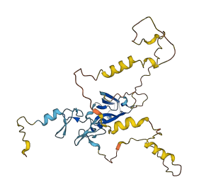
| AF-O70209-F1 | Predicted | AlphaFoldDB |
12 variants for O70209
| Variant ID(s) | Position | Change | Description | Diseaes Association | Provenance |
|---|---|---|---|---|---|
| rs3388959488 | 34 | P>L | No | EVA | |
| rs3388978914 | 37 | K>E | No | EVA | |
| rs3388959449 | 56 | G>R | No | EVA | |
| rs3388962817 | 140 | S>I | No | EVA | |
| rs32908969 | 171 | L>P | No | EVA | |
| rs36918522 | 195 | D>E | No | EVA | |
| rs230046822 | 198 | A>T | No | EVA | |
| rs3388975686 | 233 | V>D | No | EVA | |
| rs3388982015 | 235 | G>C | No | EVA | |
| rs3388986441 | 256 | A>V | No | EVA | |
| rs3388981823 | 289 | G>R | No | EVA | |
| rs3388981598 | 317 | A>K | No | EVA |
No associated diseases with O70209
1 regional properties for O70209
| Type | Name | Position | InterPro Accession |
|---|---|---|---|
| domain | Band 7 domain | 26 - 205 | IPR001107 |
6 GO annotations of cellular component
| Name | Definition |
|---|---|
| actin cytoskeleton | The part of the cytoskeleton (the internal framework of a cell) composed of actin and associated proteins. Includes actin cytoskeleton-associated complexes. |
| adherens junction | A cell-cell junction composed of the epithelial cadherin-catenin complex. The epithelial cadherins, or E-cadherins, of each interacting cell extend through the plasma membrane into the extracellular space and bind to each other. The E-cadherins bind to catenins on the cytoplasmic side of the membrane, where the E-cadherin-catenin complex binds to cytoskeletal components and regulatory and signaling molecules. |
| cytosol | The part of the cytoplasm that does not contain organelles but which does contain other particulate matter, such as protein complexes. |
| filamentous actin | A two-stranded helical polymer of the protein actin. |
| stress fiber | A contractile actin filament bundle that consists of short actin filaments with alternating polarity, cross-linked by alpha-actinin and possibly other actin bundling proteins, and with myosin present in a periodic distribution along the fiber. |
| Z disc | Platelike region of a muscle sarcomere to which the plus ends of actin filaments are attached. |
6 GO annotations of molecular function
| Name | Definition |
|---|---|
| actin binding | Binding to monomeric or multimeric forms of actin, including actin filaments. |
| actinin binding | Binding to actinin, any member of a family of proteins that crosslink F-actin. |
| cytoskeletal protein binding | Binding to a protein component of a cytoskeleton (actin, microtubule, or intermediate filament cytoskeleton). |
| metal ion binding | Binding to a metal ion. |
| muscle alpha-actinin binding | Binding to muscle isoforms of actinin. Muscle alpha-actinin isoforms are found in skeletal and cardiac muscle and are localized to the Z-disc. |
| structural constituent of muscle | The action of a molecule that contributes to the structural integrity of a muscle fiber. |
4 GO annotations of biological process
| Name | Definition |
|---|---|
| actin cytoskeleton organization | A process that is carried out at the cellular level which results in the assembly, arrangement of constituent parts, or disassembly of cytoskeletal structures comprising actin filaments and their associated proteins. |
| actin filament organization | A process that is carried out at the cellular level which results in the assembly, arrangement of constituent parts, or disassembly of cytoskeletal structures comprising actin filaments. Includes processes that control the spatial distribution of actin filaments, such as organizing filaments into meshworks, bundles, or other structures, as by cross-linking. |
| heart development | The process whose specific outcome is the progression of the heart over time, from its formation to the mature structure. The heart is a hollow, muscular organ, which, by contracting rhythmically, keeps up the circulation of the blood. |
| muscle structure development | The progression of a muscle structure over time, from its formation to its mature state. Muscle structures are contractile cells, tissues or organs that are found in multicellular organisms. |
10 homologous proteins in AiPD
| UniProt AC | Gene Name | Protein Name | Species | Evidence Code |
|---|---|---|---|---|
| Q3SYZ8 | PDLIM3 | PDZ and LIM domain protein 3 | Bos taurus (Bovine) | PR |
| P49024 | PXN | Paxillin | Gallus gallus (Chicken) | PR |
| Q9PU47 | PDLIM3 | PDZ and LIM domain protein 3 | Gallus gallus (Chicken) | PR |
| P49023 | PXN | Paxillin | Homo sapiens (Human) | PR |
| O43294 | TGFB1I1 | Transforming growth factor beta-1-induced transcript 1 protein | Homo sapiens (Human) | PR |
| Q53GG5 | PDLIM3 | PDZ and LIM domain protein 3 | Homo sapiens (Human) | PR |
| Q62219 | Tgfb1i1 | Transforming growth factor beta-1-induced transcript 1 protein | Mus musculus (Mouse) | PR |
| Q8VI36 | Pxn | Paxillin | Mus musculus (Mouse) | PR |
| Q6QGC0 | PDLIM3 | PDZ and LIM domain protein 3 | Sus scrofa (Pig) | PR |
| Q66H76 | Pxn | Paxillin | Rattus norvegicus (Rat) | PR |
| 10 | 20 | 30 | 40 | 50 | 60 |
| MPQNVVLPGP | APWGFRLSGG | IDFNQPLVIT | RITPGSKAAA | ANLCPGDVIL | AIDGFGTESM |
| 70 | 80 | 90 | 100 | 110 | 120 |
| THADAQDRIK | AASYQLCLKI | DRAETRLWSP | QVSEDGKAHP | FKINLEAEPQ | EFKPIGTAHN |
| 130 | 140 | 150 | 160 | 170 | 180 |
| RRAQPFVAAA | NIDDKRQVVS | ASYNSPIGLY | STSNIQDALH | GQLRGLIPGS | LQNEPTASVP |
| 190 | 200 | 210 | 220 | 230 | 240 |
| PQSDVYRMLH | DNRDDPAAPR | QSGSFRVLQD | LVNDGPDDRP | AGTRSVRAPV | TKVHGGAGSA |
| 250 | 260 | 270 | 280 | 290 | 300 |
| QRMPLCDKCG | SGIVGAVVKA | RDKYRHPECF | VCADCNLNLK | QKGYFFVEGE | LYCETHARAR |
| 310 | |||||
| TRPPEGYDTV | TLYPKA |