Q9QYS2
Gene name |
Grm3 (Gprc1c, Mglur3) |
Protein name |
Metabotropic glutamate receptor 3 |
Names |
mGluR3 |
Species |
Mus musculus (Mouse) |
KEGG Pathway |
mmu:108069 |
EC number |
|
Protein Class |
|
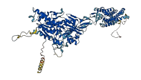
Descriptions
The autoinhibited protein was predicted that may have potential autoinhibitory elements via cis-regPred.
Autoinhibitory domains (AIDs)
Target domain |
|
Relief mechanism |
|
Assay |
cis-regPred |
Accessory elements
No accessory elements
Autoinhibited structure

Activated structure
1 structures for Q9QYS2
| Entry ID | Method | Resolution | Chain | Position | Source |
|---|---|---|---|---|---|
| AF-Q9QYS2-F1 | Predicted | AlphaFoldDB |
22 variants for Q9QYS2
| Variant ID(s) | Position | Change | Description | Diseaes Association | Provenance |
|---|---|---|---|---|---|
| rs3388748960 | 6 | R>S | No | EVA | |
| rs3388733093 | 38 | D>E | No | EVA | |
| rs3388735755 | 56 | E>V | No | EVA | |
| rs3388726819 | 59 | R>G | No | EVA | |
| rs3388744547 | 73 | L>M | No | EVA | |
| rs3388744864 | 79 | I>T | No | EVA | |
| rs3388739427 | 93 | V>G | No | EVA | |
| rs3388726823 | 112 | F>S | No | EVA | |
| rs3388737326 | 115 | A>S | No | EVA | |
| rs3388739846 | 178 | L>H | No | EVA | |
| rs3388743391 | 221 | D>E | No | EVA | |
| rs3388743345 | 222 | Y>* | No | EVA | |
| rs3388744892 | 224 | E>* | No | EVA | |
| rs3388744807 | 302 | G>D | No | EVA | |
| rs3388744860 | 373 | C>Y | No | EVA | |
| rs3388744886 | 440 | F>S | No | EVA | |
| rs49639195 | 501 | R>Q | No | EVA | |
| rs244427434 | 571 | R>K | No | EVA | |
| rs3395197342 | 608 | K>N | No | EVA | |
| rs3394911387 | 853 | Y>* | No | EVA | |
| rs3394911387 | 853 | Y>LH* | No | EVA | |
| rs3388742487 | 874 | S>P | No | EVA |
No associated diseases with Q9QYS2
No regional properties for Q9QYS2
| Type | Name | Position | InterPro Accession |
|---|---|---|---|
| No domain, repeats, and functional sites for Q9QYS2 | |||
13 GO annotations of cellular component
| Name | Definition |
|---|---|
| astrocyte projection | A prolongation or process extending from the soma of an astrocyte and wrapping around neurons. |
| axon | The long process of a neuron that conducts nerve impulses, usually away from the cell body to the terminals and varicosities, which are sites of storage and release of neurotransmitter. |
| dendritic spine | A small, membranous protrusion from a dendrite that forms a postsynaptic compartment, typically receiving input from a single presynapse. They function as partially isolated biochemical and an electrical compartments. Spine morphology is variable:they can be thin, stubby, mushroom, or branched, with a continuum of intermediate morphologies. They typically terminate in a bulb shape, linked to the dendritic shaft by a restriction. Spine remodeling is though to be involved in synaptic plasticity. |
| glutamatergic synapse | A synapse that uses glutamate as a neurotransmitter. |
| integral component of plasma membrane | The component of the plasma membrane consisting of the gene products and protein complexes having at least some part of their peptide sequence embedded in the hydrophobic region of the membrane. |
| integral component of postsynaptic membrane | The component of the postsynaptic membrane consisting of the gene products and protein complexes having at least some part of their peptide sequence embedded in the hydrophobic region of the membrane. |
| integral component of presynaptic membrane | The component of the presynaptic membrane consisting of the gene products and protein complexes having at least some part of their peptide sequence embedded in the hydrophobic region of the membrane. |
| neuron projection | A prolongation or process extending from a nerve cell, e.g. an axon or dendrite. |
| plasma membrane | The membrane surrounding a cell that separates the cell from its external environment. It consists of a phospholipid bilayer and associated proteins. |
| postsynaptic density | An electron dense network of proteins within and adjacent to the postsynaptic membrane of an asymmetric, neuron-neuron synapse. Its major components include neurotransmitter receptors and the proteins that spatially and functionally organize them such as anchoring and scaffolding molecules, signaling enzymes and cytoskeletal components. |
| postsynaptic membrane | A specialized area of membrane facing the presynaptic membrane on the tip of the nerve ending and separated from it by a minute cleft (the synaptic cleft). Neurotransmitters cross the synaptic cleft and transmit the signal to the postsynaptic membrane. |
| presynaptic active zone | A specialized region of the plasma membrane and cell cortex of a presynaptic neuron; encompasses a region of the plasma membrane where synaptic vesicles dock and fuse, and a specialized cortical cytoskeletal matrix. |
| presynaptic membrane | A specialized area of membrane of the axon terminal that faces the plasma membrane of the neuron or muscle fiber with which the axon terminal establishes a synaptic junction; many synaptic junctions exhibit structural presynaptic characteristics, such as conical, electron-dense internal protrusions, that distinguish it from the remainder of the axon plasma membrane. |
4 GO annotations of molecular function
| Name | Definition |
|---|---|
| adenylate cyclase inhibiting G protein-coupled glutamate receptor activity | Combining with glutamate and transmitting the signal across the membrane by activating the alpha-subunit of an associated heterotrimeric G-protein complex to inhibit downstream adenylate cyclase activity. |
| calcium channel regulator activity | Modulates the activity of a calcium channel. |
| group II metabotropic glutamate receptor activity | A G protein-coupled receptor that is activated by trans-1-aminocyclopentane-1,3-dicarboxylic acid (t-ACPD) and inhibits adenylate cyclase activity. |
| scaffold protein binding | Binding to a scaffold protein. Scaffold proteins are crucial regulators of many key signaling pathways. Although not strictly defined in function, they are known to interact and/or bind with multiple members of a signaling pathway, tethering them into complexes. |
7 GO annotations of biological process
| Name | Definition |
|---|---|
| G protein-coupled glutamate receptor signaling pathway | A G protein-coupled receptor signaling pathway initiated by glutamate binding to its receptor on the surface of a target cell, and ending with the regulation of a downstream cellular process. |
| gene expression | The process in which a gene's sequence is converted into a mature gene product (protein or RNA). This includes the production of an RNA transcript and its processing, translation and maturation for protein-coding genes. |
| modulation of chemical synaptic transmission | Any process that modulates the frequency or amplitude of synaptic transmission, the process of communication from a neuron to a target (neuron, muscle, or secretory cell) across a synapse. Amplitude, in this case, refers to the change in postsynaptic membrane potential due to a single instance of synaptic transmission. |
| regulation of sensory perception of pain | Any process that modulates the frequency, rate or extent of the sensory perception of pain, the series of events required for an organism to receive a painful stimulus, convert it to a molecular signal, and recognize and characterize the signal. |
| regulation of synaptic transmission, glutamatergic | Any process that modulates the frequency, rate or extent of glutamatergic synaptic transmission, the process of communication from a neuron to another neuron across a synapse using the neurotransmitter glutamate. |
| sensory perception of pain | The series of events required for an organism to receive a painful stimulus, convert it to a molecular signal, and recognize and characterize the signal. Pain is medically defined as the physical sensation of discomfort or distress caused by injury or illness, so can hence be described as a harmful stimulus which signals current (or impending) tissue damage. Pain may come from extremes of temperature, mechanical damage, electricity or from noxious chemical substances. This is a neurological process. |
| synaptic transmission, glutamatergic | The vesicular release of glutamate from a presynapse, across a chemical synapse, the subsequent activation of glutamate receptors at the postsynapse of a target cell (neuron, muscle, or secretory cell) and the effects of this activation on the postsynaptic membrane potential and ionic composition of the postsynaptic cytosol. This process encompasses both spontaneous and evoked release of neurotransmitter and all parts of synaptic vesicle exocytosis. Evoked transmission starts with the arrival of an action potential at the presynapse. |
8 homologous proteins in AiPD
| UniProt AC | Gene Name | Protein Name | Species | Evidence Code |
|---|---|---|---|---|
| O00222 | GRM8 | Metabotropic glutamate receptor 8 | Homo sapiens (Human) | PR |
| Q14416 | GRM2 | Metabotropic glutamate receptor 2 | Homo sapiens (Human) | PR |
| P41180 | CASR | Extracellular calcium-sensing receptor | Homo sapiens (Human) | PR |
| P47743 | Grm8 | Metabotropic glutamate receptor 8 | Mus musculus (Mouse) | PR |
| Q14BI2 | Grm2 | Metabotropic glutamate receptor 2 | Mus musculus (Mouse) | PR |
| Q3UVX5 | Grm5 | Metabotropic glutamate receptor 5 | Mus musculus (Mouse) | PR |
| P31421 | Grm2 | Metabotropic glutamate receptor 2 | Rattus norvegicus (Rat) | PR |
| P31422 | Grm3 | Metabotropic glutamate receptor 3 | Rattus norvegicus (Rat) | PR |
| 10 | 20 | 30 | 40 | 50 | 60 |
| MKMLTRLQVL | MLALFSKGFL | VSLGDHNFMR | REIKIEGDLV | LGGLFPINEK | GTGTEECGRI |
| 70 | 80 | 90 | 100 | 110 | 120 |
| NEDRGIQRLE | AMLFAIDEIN | KDNYLLPGVK | LGVHILDTCS | RDTYALEQSL | EFVRASLTKV |
| 130 | 140 | 150 | 160 | 170 | 180 |
| DEAEYMCPDG | SYAIQENIPL | LIAGVIGGSY | SSVSIQVANL | LRLFQIPQIS | YASTSAKLSD |
| 190 | 200 | 210 | 220 | 230 | 240 |
| KSRYDYFART | VPPDFYQAKA | MAEILRYFNW | TYVSTVASEG | DYGETGIEAF | EQEARLRNIC |
| 250 | 260 | 270 | 280 | 290 | 300 |
| IATAEKVGRS | NIRKSYDSVI | RELLQKPNAR | VVVLFMRSDD | SRELIAAASR | VNASFTWVAS |
| 310 | 320 | 330 | 340 | 350 | 360 |
| DGWGAQESIV | KGSEHVAYGA | ITLELASHPV | RQFDRYFQSL | NPYNNHRNPW | FRDFWEQKFQ |
| 370 | 380 | 390 | 400 | 410 | 420 |
| CSLQNKRNHR | QICDKHLAID | SSNYEQESKI | MFVVNAVYAM | AHALHKMQRT | LCPNTTKLCD |
| 430 | 440 | 450 | 460 | 470 | 480 |
| AMKILDGKKL | YKDYLLKINF | TAPFNPNKGA | DSIVKFDTYG | DGMGRYNVFN | FQHIGGKYSY |
| 490 | 500 | 510 | 520 | 530 | 540 |
| LKVGHWAETL | YLDVDSIHWS | RNSVPTSQCS | DPCAPNEMKN | MQPGDVCCWI | CIPCEPYEYL |
| 550 | 560 | 570 | 580 | 590 | 600 |
| VDEFTCMDCG | PGQWPTADLS | GCYNLPEDYI | RWEDAWAIGP | VTIACLGFMC | TCIVITVFIK |
| 610 | 620 | 630 | 640 | 650 | 660 |
| HNNTPLVKAS | GRELCYILLF | GVSLSYCMTF | FFIAKPSPVI | CALRRLGLGT | SFAICYSALL |
| 670 | 680 | 690 | 700 | 710 | 720 |
| TKTNCIARIF | DGVKNGAQRP | KFISPSSQVF | ICLGLILVQI | VMVSVWLILE | TPGTRRYTLP |
| 730 | 740 | 750 | 760 | 770 | 780 |
| EKRETVILKC | NVKDSSMLIS | LTYDVVLVIL | CTVYAFKTRK | CPENFNEAKF | IGFTMYTTCI |
| 790 | 800 | 810 | 820 | 830 | 840 |
| IWLAFLPIFY | VTSSDYRVQT | TTMCISVSLS | GFVVLGCLFA | PKVHIVLFQP | QKNVVTHRLH |
| 850 | 860 | 870 | |||
| LNRFSVSGTA | TTYSQSSAST | YVPTVCNGRE | VLDSTTSSL |