Q28175
Gene name |
RPE65 |
Protein name |
Retinoid isomerohydrolase |
Names |
All-trans-retinyl-palmitate hydrolase, Lutein isomerase, Meso-zeaxanthin isomerase, Retinal pigment epithelium-specific 65 kDa protein, Retinol isomerase |
Species |
Bos taurus (Bovine) |
KEGG Pathway |
bta:282043 |
EC number |
3.1.1.64: Carboxylic ester hydrolases |
Protein Class |
|
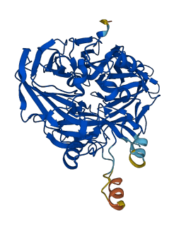
Descriptions
The autoinhibited protein was predicted that may have potential autoinhibitory elements via cis-regPred.
Autoinhibitory domains (AIDs)
Target domain |
|
Relief mechanism |
|
Assay |
cis-regPred |
Accessory elements
No accessory elements
Autoinhibited structure

Activated structure
20 structures for Q28175
| Entry ID | Method | Resolution | Chain | Position | Source |
|---|---|---|---|---|---|
| 3FSN | X-ray | 214 A | A/B | 1-533 | PDB |
| 3KVC | X-ray | 190 A | A/B | 1-533 | PDB |
| 4F2Z | X-ray | 300 A | A/E | 1-533 | PDB |
| 4F30 | X-ray | 315 A | A | 1-533 | PDB |
| 4F3A | X-ray | 260 A | A | 1-533 | PDB |
| 4F3D | X-ray | 250 A | A/B | 1-533 | PDB |
| 4RSC | X-ray | 180 A | A/B | 1-533 | PDB |
| 4RSE | X-ray | 239 A | A/B | 2-533 | PDB |
| 4RYX | X-ray | 200 A | A | 1-533 | PDB |
| 4RYY | X-ray | 230 A | A/B | 2-533 | PDB |
| 4RYZ | X-ray | 250 A | A/B | 2-533 | PDB |
| 4ZHK | X-ray | 209 A | A/B | 2-533 | PDB |
| 5UL5 | X-ray | 220 A | A/B | 1-533 | PDB |
| 5ULG | X-ray | 210 A | A/B | 1-533 | PDB |
| 7K88 | X-ray | 210 A | A/B | 2-533 | PDB |
| 7K89 | X-ray | 215 A | A/B | 2-533 | PDB |
| 7K8G | X-ray | 195 A | A/B | 2-533 | PDB |
| 7L0E | X-ray | 190 A | A/B | 1-533 | PDB |
| 8DOC | X-ray | 210 A | A | 2-533 | PDB |
| AF-Q28175-F1 | Predicted | AlphaFoldDB |
No variants for Q28175
| Variant ID(s) | Position | Change | Description | Diseaes Association | Provenance |
|---|---|---|---|---|---|
| No variants for Q28175 | |||||
No associated diseases with Q28175
Functions
| Description | ||
|---|---|---|
| EC Number | 3.1.1.64 | Carboxylic ester hydrolases |
| Subcellular Localization |
|
|
| PANTHER Family | ||
| PANTHER Subfamily | ||
| PANTHER Protein Class | ||
| PANTHER Pathway Category | No pathway information available | |
3 GO annotations of cellular component
| Name | Definition |
|---|---|
| endoplasmic reticulum membrane | The lipid bilayer surrounding the endoplasmic reticulum. |
| membrane | A lipid bilayer along with all the proteins and protein complexes embedded in it an attached to it. |
| plasma membrane | The membrane surrounding a cell that separates the cell from its external environment. It consists of a phospholipid bilayer and associated proteins. |
9 GO annotations of molecular function
| Name | Definition |
|---|---|
| all-trans-retinyl-ester hydrolase, 11-cis retinol forming activity | Catalysis of the reaction: H(2)O + all-trans-retinyl ester = 11-cis-retinol + fatty acid. |
| all-trans-retinyl-palmitate hydrolase, 11-cis retinol forming activity | Catalysis of the reaction: H(2)O + all-trans-retinyl palmitate = 11-cis-retinol + H(+) + palmitate. |
| cardiolipin binding | Binding to cardiolipin. |
| identical protein binding | Binding to an identical protein or proteins. |
| metal ion binding | Binding to a metal ion. |
| oxidoreductase activity, acting on single donors with incorporation of molecular oxygen, incorporation of two atoms of oxygen | Catalysis of an oxidation-reduction (redox) reaction in which hydrogen or electrons are transferred from one donor, and two oxygen atoms is incorporated into a donor. |
| phosphatidylcholine binding | Binding to a phosphatidylcholine, a glycophospholipid in which a phosphatidyl group is esterified to the hydroxyl group of choline. |
| phosphatidylserine binding | Binding to phosphatidylserine, a class of glycophospholipids in which a phosphatidyl group is esterified to the hydroxyl group of L-serine. |
| retinal isomerase activity | Catalysis of the reaction: all-trans-retinal = 11-cis-retinal. |
3 GO annotations of biological process
| Name | Definition |
|---|---|
| response to stimulus | Any process that results in a change in state or activity of a cell or an organism (in terms of movement, secretion, enzyme production, gene expression, etc.) as a result of a stimulus. The process begins with detection of the stimulus and ends with a change in state or activity or the cell or organism. |
| visual perception | The series of events required for an organism to receive a visual stimulus, convert it to a molecular signal, and recognize and characterize the signal. Visual stimuli are detected in the form of photons and are processed to form an image. |
| zeaxanthin biosynthetic process | The chemical reactions and pathways resulting in the formation of zeaxanthin. |
10 homologous proteins in AiPD
| UniProt AC | Gene Name | Protein Name | Species | Evidence Code |
|---|---|---|---|---|
| Q9YGX2 | RPE65 | Retinoid isomerohydrolase | Gallus gallus (Chicken) | PR |
| Q9TVB8 | RPE65 | Retinoid isomerohydrolase | Canis lupus familiaris (Dog) (Canis familiaris) | PR |
| Q16518 | RPE65 | Retinoid isomerohydrolase | Homo sapiens (Human) | PR |
| O24592 | VP14 | 9-cis-epoxycarotenoid dioxygenase 1, chloroplastic | Zea mays (Maize) | PR |
| Q91ZQ5 | Rpe65 | Retinoid isomerohydrolase | Mus musculus (Mouse) | PR |
| O70276 | Rpe65 | Retinoid isomerohydrolase | Rattus norvegicus (Rat) | PR |
| Q8LIY8 | CCD8B | Carotenoid cleavage dioxygenase 8 homolog B, chloroplastic | Oryza sativa subsp japonica (Rice) | PR |
| Q9C6Z1 | NCED5 | Probable 9-cis-epoxycarotenoid dioxygenase NCED5, chloroplastic | Arabidopsis thaliana (Mouse-ear cress) | PR |
| Q9LRR7 | NCED3 | 9-cis-epoxycarotenoid dioxygenase NCED3, chloroplastic | Arabidopsis thaliana (Mouse-ear cress) | PR |
| Q9LRM7 | NCED6 | 9-cis-epoxycarotenoid dioxygenase NCED6, chloroplastic | Arabidopsis thaliana (Mouse-ear cress) | PR |
| 10 | 20 | 30 | 40 | 50 | 60 |
| MSSQVEHPAG | GYKKLFETVE | ELSSPLTAHV | TGRIPLWLTG | SLLRCGPGLF | EVGSEPFYHL |
| 70 | 80 | 90 | 100 | 110 | 120 |
| FDGQALLHKF | DFKEGHVTYH | RRFIRTDAYV | RAMTEKRIVI | TEFGTCAFPD | PCKNIFSRFF |
| 130 | 140 | 150 | 160 | 170 | 180 |
| SYFRGVEVTD | NALVNIYPVG | EDYYACTETN | FITKVNPETL | ETIKQVDLCN | YVSVNGATAH |
| 190 | 200 | 210 | 220 | 230 | 240 |
| PHIENDGTVY | NIGNCFGKNF | SIAYNIVKIP | PLQADKEDPI | SKSEIVVQFP | CSDRFKPSYV |
| 250 | 260 | 270 | 280 | 290 | 300 |
| HSFGLTPNYI | VFVETPVKIN | LFKFLSSWSL | WGANYMDCFE | SNETMGVWLH | IADKKRKKYI |
| 310 | 320 | 330 | 340 | 350 | 360 |
| NNKYRTSPFN | LFHHINTYED | HEFLIVDLCC | WKGFEFVYNY | SYLANLRENW | EEVKKNARKA |
| 370 | 380 | 390 | 400 | 410 | 420 |
| PQPEVRRYVL | PLNIDKADTG | KNLVTLPNTT | ATAILCSDET | IWLEPEVLFS | GPRQAFEFPQ |
| 430 | 440 | 450 | 460 | 470 | 480 |
| INYQKYGGKP | YTYAYGLGLN | HFVPDRLCKL | NVKTKETWVW | QEPDSYPSEP | IFVSHPDALE |
| 490 | 500 | 510 | 520 | 530 | |
| EDDGVVLSVV | VSPGAGQKPA | YLLILNAKDL | SEVARAEVEI | NIPVTFHGLF | KKS |