Q9Z2C5
Gene name |
Mtm1 |
Protein name |
Myotubularin |
Names |
Phosphatidylinositol-3,5-bisphosphate 3-phosphatase, Phosphatidylinositol-3-phosphate phosphatase |
Species |
Mus musculus (Mouse) |
KEGG Pathway |
mmu:17772 |
EC number |
3.1.3.64: Phosphoric monoester hydrolases |
Protein Class |
|
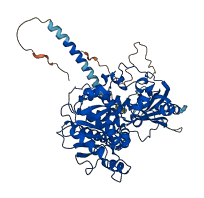
Descriptions
The autoinhibited protein was predicted that may have potential autoinhibitory elements via cis-regPred.
Autoinhibitory domains (AIDs)
Target domain |
|
Relief mechanism |
|
Assay |
cis-regPred |
Accessory elements
No accessory elements
Autoinhibited structure

Activated structure
1 structures for Q9Z2C5
| Entry ID | Method | Resolution | Chain | Position | Source |
|---|---|---|---|---|---|
| AF-Q9Z2C5-F1 | Predicted | AlphaFoldDB |
36 variants for Q9Z2C5
| Variant ID(s) | Position | Change | Description | Diseaes Association | Provenance |
|---|---|---|---|---|---|
| rs3389558080 | 3 | S>F | No | EVA | |
| rs3410505119 | 37 | R>Q | No | EVA | |
| rs233060719 | 43 | L>P | No | EVA | |
| rs3410659995 | 46 | E>D | No | EVA | |
| rs1134563859 | 97 | G>V | No | EVA | |
| rs1133542676 | 99 | A>T | No | EVA | |
| rs1134304703 | 100 | T>I | No | EVA | |
| rs3411240467 | 152 | A>T | No | EVA | |
| rs3409361874 | 153 | F>I | No | EVA | |
| rs3409361821 | 153 | F>Y | No | EVA | |
| rs3410819570 | 154 | V>A | No | EVA | |
| rs3410506508 | 156 | E>K | No | EVA | |
| rs3389577330 | 164 | W>* | No | EVA | |
| rs3389555222 | 183 | W>* | No | EVA | |
| rs3389555222 | 183 | W>S | No | EVA | |
| rs3389574773 | 196 | E>G | No | EVA | |
| rs31685040 | 218 | T>M | No | EVA | |
| rs3409811058 | 228 | L>V | No | EVA | |
| rs3410871738 | 286 | V>L | No | EVA | |
| rs3409276419 | 309 | L>F | No | EVA | |
| rs3412670577 | 313 | N>K | No | EVA | |
| rs3389529140 | 351 | K>N | No | EVA | |
| rs3389555198 | 375 | C>R | No | EVA | |
| rs3409361899 | 418 | F>I | No | EVA | |
| rs3410820887 | 426 | D>V | No | EVA | |
| rs3410506498 | 426 | D>Y | No | EVA | |
| rs3410765528 | 427 | K>* | No | EVA | |
| rs3410506509 | 427 | K>N | No | EVA | |
| rs3410506485 | 428 | N>H | No | EVA | |
| rs3389574756 | 437 | I>N | No | EVA | |
| rs3389541943 | 446 | W>L | No | EVA | |
| rs3389555166 | 461 | G>C | No | EVA | |
| rs3410765607 | 496 | V>A | No | EVA | |
| rs3389557029 | 511 | N>D | No | EVA | |
| rs3389558085 | 538 | Y>C | No | EVA | |
| rs3389518426 | 564 | R>H | No | EVA |
No associated diseases with Q9Z2C5
5 regional properties for Q9Z2C5
| Type | Name | Position | InterPro Accession |
|---|---|---|---|
| domain | Tyrosine-specific protein phosphatases domain | 386 - 433 | IPR000387 |
| domain | Protein-tyrosine phosphatase, catalytic | 365 - 513 | IPR003595 |
| domain | GRAM domain | 65 - 185 | IPR004182 |
| domain | Myotubularin-like, phosphatase domain | 192 - 580 | IPR010569 |
| active_site | Protein-tyrosine phosphatase, active site | 415 - 425 | IPR016130 |
Functions
| Description | ||
|---|---|---|
| EC Number | 3.1.3.64 | Phosphoric monoester hydrolases |
| Subcellular Localization |
|
|
| PANTHER Family | ||
| PANTHER Subfamily | ||
| PANTHER Protein Class | ||
| PANTHER Pathway Category | No pathway information available | |
7 GO annotations of cellular component
| Name | Definition |
|---|---|
| cytoplasm | The contents of a cell excluding the plasma membrane and nucleus, but including other subcellular structures. |
| cytosol | The part of the cytoplasm that does not contain organelles but which does contain other particulate matter, such as protein complexes. |
| filopodium | Thin, stiff, actin-based protrusion extended by the leading edge of a motile cell such as a crawling fibroblast or amoeba, or an axonal or dendritic growth cone, or a dendritic shaft. |
| I band | A region of a sarcomere that appears as a light band on each side of the Z disc, comprising a region of the sarcomere where thin (actin) filaments are not overlapped by thick (myosin) filaments; contains actin, troponin, and tropomyosin; each sarcomere includes half of an I band at each end. |
| late endosome | A prelysosomal endocytic organelle differentiated from early endosomes by lower lumenal pH and different protein composition. Late endosomes are more spherical than early endosomes and are mostly juxtanuclear, being concentrated near the microtubule organizing center. |
| plasma membrane | The membrane surrounding a cell that separates the cell from its external environment. It consists of a phospholipid bilayer and associated proteins. |
| ruffle | Projection at the leading edge of a crawling cell; the protrusions are supported by a microfilament meshwork. |
5 GO annotations of molecular function
| Name | Definition |
|---|---|
| intermediate filament binding | Binding to an intermediate filament, a distinct elongated structure, characteristically 10 nm in diameter, that occurs in the cytoplasm of higher eukaryotic cells. Intermediate filaments form a fibrous system, composed of chemically heterogeneous subunits and involved in mechanically integrating the various components of the cytoplasmic space. |
| phosphatidylinositol binding | Binding to an inositol-containing glycerophospholipid, i.e. phosphatidylinositol (PtdIns) and its phosphorylated derivatives. |
| phosphatidylinositol-3,5-bisphosphate 3-phosphatase activity | Catalysis of the reaction: 1-phosphatidyl-1D-myo-inositol 3,5-bisphosphate + H2O = a 1-phosphatidyl-1D-myo-inositol 5-phosphate + phosphate + 2 H+. |
| phosphatidylinositol-3-phosphatase activity | Catalysis of the reaction: 1-phosphatidyl-1D-myo-inositol 3-phosphate + H2O = 1-phosphatidyl-1D-myo-inositol + phosphate. |
| phosphoprotein phosphatase activity | Catalysis of the reaction: a phosphoprotein + H2O = a protein + phosphate. Together with protein kinases, these enzymes control the state of phosphorylation of cellular proteins and thereby provide an important mechanism for regulating cellular activity. |
19 GO annotations of biological process
| Name | Definition |
|---|---|
| autophagosome assembly | The formation of a double membrane-bounded structure, the autophagosome, that occurs when a specialized membrane sac, called the isolation membrane, starts to enclose a portion of the cytoplasm. |
| endosome to lysosome transport | The directed movement of substances from endosomes to lysosomes. |
| intermediate filament organization | Control of the spatial distribution of intermediate filaments; includes organizing filaments into meshworks, bundles, or other structures, as by cross-linking. |
| mitochondrion distribution | Any process that establishes the spatial arrangement of mitochondria between and within cells. |
| mitochondrion morphogenesis | The process in which the anatomical structures of a mitochondrion are generated and organized. |
| muscle cell cellular homeostasis | The cellular homeostatic process that preserves a muscle cell in a stable functional or structural state. |
| negative regulation of autophagosome assembly | Any process that stops, prevents or reduces the frequency, rate or extent of autophagosome assembly. |
| negative regulation of proteasomal ubiquitin-dependent protein catabolic process | Any process that stops, prevents, or reduces the frequency, rate or extent of the breakdown of a protein or peptide by hydrolysis of its peptide bonds, initiated by the covalent attachment of ubiquitin, and mediated by the proteasome. |
| negative regulation of protein kinase B signaling | Any process that stops, prevents, or reduces the frequency, rate or extent of protein kinase B signaling, a series of reactions mediated by the intracellular serine/threonine kinase protein kinase B. |
| negative regulation of TOR signaling | Any process that stops, prevents, or reduces the frequency, rate or extent of TOR signaling. |
| phosphatidylinositol dephosphorylation | The process of removing one or more phosphate groups from a phosphatidylinositol. |
| positive regulation of skeletal muscle tissue growth | Any process that activates, maintains or increases the rate of skeletal muscle growth. |
| proteasome-mediated ubiquitin-dependent protein catabolic process | The chemical reactions and pathways resulting in the breakdown of a protein or peptide by hydrolysis of its peptide bonds, initiated by the covalent attachment of ubiquitin, and mediated by the proteasome. |
| protein dephosphorylation | The process of removing one or more phosphoric residues from a protein. |
| protein kinase B signaling | A series of reactions, mediated by the intracellular serine/threonine kinase protein kinase B (also called AKT), which occurs as a result of a single trigger reaction or compound. |
| protein transport | The directed movement of proteins into, out of or within a cell, or between cells, by means of some agent such as a transporter or pore. |
| regulation of vacuole organization | Any process that modulates the frequency, rate or extent of a process involved in the formation, arrangement of constituent parts, or disassembly of a vacuole. |
| skeletal muscle tissue growth | The increase in size or mass of a skeletal muscle. This may be due to a change in the fiber number or size. |
| TOR signaling | The series of molecular signals mediated by TOR (Target of rapamycin) proteins, members of the phosphoinositide (PI) 3-kinase related kinase (PIKK) family that act as serine/threonine kinases in response to nutrient availability or growth factors. |
6 homologous proteins in AiPD
| UniProt AC | Gene Name | Protein Name | Species | Evidence Code |
|---|---|---|---|---|
| A6QLT4 | MTM1 | Myotubularin | Bos taurus (Bovine) | PR |
| Q13496 | MTM1 | Myotubularin | Homo sapiens (Human) | PR |
| Q9Z2C4 | Mtmr1 | Myotubularin-related protein 1 | Mus musculus (Mouse) | PR |
| Q9Z2D1 | Mtmr2 | Myotubularin-related protein 2 | Mus musculus (Mouse) | PR |
| Q9Z2C9 | Mtmr7 | Myotubularin-related protein 7 | Mus musculus (Mouse) | PR |
| Q5EB32 | mtm1 | Myotubularin | Xenopus tropicalis (Western clawed frog) (Silurana tropicalis) | PR |
| 10 | 20 | 30 | 40 | 50 | 60 |
| MASASASKYN | SHSLENESIK | KVSQDGVSQD | VSETVPRLPG | ELLITEKEVI | YICPFNGPIK |
| 70 | 80 | 90 | 100 | 110 | 120 |
| GRVYITNYRL | YLRSLETDSA | LILDVPLGVI | SRIEKMGGAT | SRGENSYGLD | ITCKDLRNLR |
| 130 | 140 | 150 | 160 | 170 | 180 |
| FALKQEGHSR | RDMFEILVKH | AFPLAHNLPL | FAFVNEEKFN | VDGWTVYNPV | EEYRRQGLPN |
| 190 | 200 | 210 | 220 | 230 | 240 |
| HHWRISFINK | CYELCETYPA | LLVVPYRTSD | DDLRRIATFR | SRNRLPVLSW | IHPENKMVIM |
| 250 | 260 | 270 | 280 | 290 | 300 |
| RCSQPLVGMS | GKRNKDDEKY | LDVIRETNKQ | TSKLMIYDAR | PSVNAVANKA | TGGGYESDDA |
| 310 | 320 | 330 | 340 | 350 | 360 |
| YQNSELSFLD | IHNIHVMRES | LKKVKDIVYP | NIEESHWLSS | LESTHWLEHI | KLVLTGAIQV |
| 370 | 380 | 390 | 400 | 410 | 420 |
| ADQVSSGKSS | VLVHCSDGWD | RTAQLTSLAM | LMLDSFYRTI | EGFEILVQKE | WISFGHKFAS |
| 430 | 440 | 450 | 460 | 470 | 480 |
| RIGHGDKNHA | DADRSPIFLQ | FIDCVWQMSK | QFPTAFEFNE | GFLITVLDHL | YSCRFGTFLF |
| 490 | 500 | 510 | 520 | 530 | 540 |
| NCDSARERQK | LTERTVSLWS | LINSNKDKFK | NPFYTKEINR | VLYPVASMRH | LELWVNYYIR |
| 550 | 560 | 570 | 580 | 590 | 600 |
| WNPRVKQQQP | NPVEQRYMEL | LALRDDYIKR | LEELQLANSA | KLADAPASTS | SSSQMVPHVQ |
| THF |