P51667
Gene name |
Myl2 |
Protein name |
Myosin regulatory light chain 2, ventricular/cardiac muscle isoform |
Names |
MLC-2, MLC-2v, Myosin light chain 2, slow skeletal/ventricular muscle isoform, MLC-2s/v |
Species |
Mus musculus (Mouse) |
KEGG Pathway |
mmu:17906 |
EC number |
|
Protein Class |
|
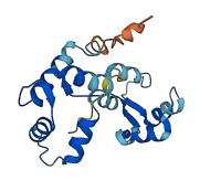
Descriptions
The autoinhibited protein was predicted that may have potential autoinhibitory elements via cis-regPred.
Autoinhibitory domains (AIDs)
Target domain |
|
Relief mechanism |
|
Assay |
cis-regPred |
Accessory elements
No accessory elements
Autoinhibited structure

Activated structure
2 structures for P51667
| Entry ID | Method | Resolution | Chain | Position | Source |
|---|---|---|---|---|---|
| 8Q6T | EM | 1800 A | E/F/L/M/T/U | 7-166 | PDB |
| AF-P51667-F1 | Predicted | AlphaFoldDB |
7 variants for P51667
| Variant ID(s) | Position | Change | Description | Diseaes Association | Provenance |
|---|---|---|---|---|---|
| rs3388788658 | 19 | S>A | No | EVA | |
| rs32209032 | 24 | T>A | No | EVA | |
| rs3388782684 | 53 | F>C | No | EVA | |
| rs3388770412 | 83 | L>P | No | EVA | |
| rs3388788595 | 85 | M>I | No | EVA | |
| rs3395753322 | 91 | K>* | No | EVA | |
| rs3388789295 | 95 | P>S | No | EVA |
No associated diseases with P51667
4 regional properties for P51667
| Type | Name | Position | InterPro Accession |
|---|---|---|---|
| domain | EF-hand domain | 24 - 73 | IPR002048-1 |
| domain | EF-hand domain | 94 - 129 | IPR002048-2 |
| domain | EF-hand domain | 134 - 162 | IPR002048-3 |
| binding_site | EF-Hand 1, calcium-binding site | 37 - 49 | IPR018247 |
8 GO annotations of cellular component
| Name | Definition |
|---|---|
| A band | The dark-staining region of a sarcomere, in which myosin thick filaments are present; the center is traversed by the paler H zone, which in turn contains the M line. |
| actin cytoskeleton | The part of the cytoskeleton (the internal framework of a cell) composed of actin and associated proteins. Includes actin cytoskeleton-associated complexes. |
| cardiac myofibril | A cardiac myofibril is a myofibril specific to cardiac muscle cells. |
| contractile fiber | Fibers, composed of actin, myosin, and associated proteins, found in cells of smooth or striated muscle. |
| cytoplasm | The contents of a cell excluding the plasma membrane and nucleus, but including other subcellular structures. |
| cytoskeleton | A cellular structure that forms the internal framework of eukaryotic and prokaryotic cells. The cytoskeleton includes intermediate filaments, microfilaments, microtubules, the microtrabecular lattice, and other structures characterized by a polymeric filamentous nature and long-range order within the cell. The various elements of the cytoskeleton not only serve in the maintenance of cellular shape but also have roles in other cellular functions, including cellular movement, cell division, endocytosis, and movement of organelles. |
| myofibril | The contractile element of skeletal and cardiac muscle; a long, highly organized bundle of actin, myosin, and other proteins that contracts by a sliding filament mechanism. |
| myosin complex | A protein complex, formed of one or more myosin heavy chains plus associated light chains and other proteins, that functions as a molecular motor; uses the energy of ATP hydrolysis to move actin filaments or to move vesicles or other cargo on fixed actin filaments; has magnesium-ATPase activity and binds actin. Myosin classes are distinguished based on sequence features of the motor, or head, domain, but also have distinct tail regions that are believed to bind specific cargoes. |
3 GO annotations of molecular function
| Name | Definition |
|---|---|
| actin monomer binding | Binding to monomeric actin, also known as G-actin. |
| calcium ion binding | Binding to a calcium ion (Ca2+). |
| structural constituent of muscle | The action of a molecule that contributes to the structural integrity of a muscle fiber. |
12 GO annotations of biological process
| Name | Definition |
|---|---|
| cardiac muscle contraction | Muscle contraction of cardiac muscle tissue. |
| cardiac myofibril assembly | The process whose specific outcome is the progression of the cardiac myofibril over time, from its formation to the mature structure. A cardiac myofibril is a myofibril specific to cardiac muscle cells. |
| heart contraction | The multicellular organismal process in which the heart decreases in volume in a characteristic way to propel blood through the body. |
| heart development | The process whose specific outcome is the progression of the heart over time, from its formation to the mature structure. The heart is a hollow, muscular organ, which, by contracting rhythmically, keeps up the circulation of the blood. |
| heart morphogenesis | The developmental process in which the heart is generated and organized. The heart is a hollow, muscular organ, which, by contracting rhythmically, keeps up the circulation of the blood. |
| muscle cell development | The process whose specific outcome is the progression of a muscle cell over time, from its formation to the mature structure. Muscle cell development does not include the steps involved in committing an unspecified cell to the muscle cell fate. |
| muscle cell fate specification | The process in which a cell becomes capable of differentiating autonomously into a muscle cell in an environment that is neutral with respect to the developmental pathway; upon specification, the cell fate can be reversed. |
| negative regulation of cell growth | Any process that stops, prevents, or reduces the frequency, rate, extent or direction of cell growth. |
| positive regulation of the force of heart contraction | Any process that increases the force of heart muscle contraction. |
| post-embryonic development | The process whose specific outcome is the progression of the organism over time, from the completion of embryonic development to the mature structure. See embryonic development. |
| regulation of the force of heart contraction | Any process that modulates the extent of heart contraction, changing the force with which blood is propelled. |
| ventricular cardiac muscle tissue morphogenesis | The process in which the anatomical structures of cardiac ventricle muscle is generated and organized. |
9 homologous proteins in AiPD
| UniProt AC | Gene Name | Protein Name | Species | Evidence Code |
|---|---|---|---|---|
| Q5E9E2 | MYL9 | Myosin regulatory light polypeptide 9 | Bos taurus (Bovine) | PR |
| P19105 | MYL12A | Myosin regulatory light chain 12A | Homo sapiens (Human) | PR |
| Q9BUA6 | MYL10 | Myosin regulatory light chain 10 | Homo sapiens (Human) | PR |
| O14950 | MYL12B | Myosin regulatory light chain 12B | Homo sapiens (Human) | PR |
| P97457 | Myl11 | Myosin regulatory light chain 11 | Mus musculus (Mouse) | PR |
| Q3THE2 | Myl12b | Myosin regulatory light chain 12B | Mus musculus (Mouse) | PR |
| Q62082 | Myl10 | Myosin regulatory light chain 10 | Mus musculus (Mouse) | PR |
| P13832 | Rlc-a | Myosin regulatory light chain RLC-A | Rattus norvegicus (Rat) | PR |
| P18666 | Myl12b | Myosin regulatory light chain 12B | Rattus norvegicus (Rat) | PR |
| 10 | 20 | 30 | 40 | 50 | 60 |
| MAPKKAKKRI | EGGSSNVFSM | FEQTQIQEFK | EAFTIMDQNR | DGFIDKNDLR | DTFAALGRVN |
| 70 | 80 | 90 | 100 | 110 | 120 |
| VKNEEIDEMI | KEAPGPINFT | VFLTMFGEKL | KGADPEETIL | NAFKVFDPEG | KGSLKADYVR |
| 130 | 140 | 150 | 160 | ||
| EMLTTQAERF | SKEEIDQMFA | AFPPDVTGNL | DYKNLVHIIT | HGEEKD |