Q3TY86
Gene name |
Aifm3 (Aifl) |
Protein name |
Apoptosis-inducing factor 3 |
Names |
Apoptosis-inducing factor-like protein |
Species |
Mus musculus (Mouse) |
KEGG Pathway |
mmu:72168 |
EC number |
|
Protein Class |
|
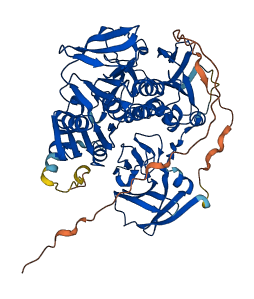
Descriptions
The autoinhibited protein was predicted that may have potential autoinhibitory elements via cis-regPred.
Autoinhibitory domains (AIDs)
Target domain |
|
Relief mechanism |
|
Assay |
cis-regPred |
Accessory elements
No accessory elements
Autoinhibited structure

Activated structure
1 structures for Q3TY86
| Entry ID | Method | Resolution | Chain | Position | Source |
|---|---|---|---|---|---|
| AF-Q3TY86-F1 | Predicted | AlphaFoldDB |
27 variants for Q3TY86
| Variant ID(s) | Position | Change | Description | Diseaes Association | Provenance |
|---|---|---|---|---|---|
| rs3389418737 | 9 | K>N | No | EVA | |
| rs3389365289 | 46 | A>S | No | EVA | |
| rs3389401178 | 108 | K>E | No | EVA | |
| rs3389396310 | 118 | K>* | No | EVA | |
| rs3389413267 | 119 | G>C | No | EVA | |
| rs3389404494 | 203 | G>D | No | EVA | |
| rs3389404561 | 243 | D>E | No | EVA | |
| rs3389407298 | 253 | P>L | No | EVA | |
| rs3389400790 | 300 | S>T | No | EVA | |
| rs3389398547 | 324 | N>D | No | EVA | |
| rs3389395035 | 337 | V>I | No | EVA | |
| rs3389396328 | 342 | F>Y | No | EVA | |
| rs3389404558 | 375 | R>C | No | EVA | |
| rs3389384410 | 392 | Y>N | No | EVA | |
| rs3406552316 | 407 | L>P | No | EVA | |
| rs3406381964 | 409 | E>D | No | EVA | |
| rs3406131409 | 411 | V>G | No | EVA | |
| rs3389398514 | 415 | S>N | No | EVA | |
| rs3389407330 | 434 | G>S | No | EVA | |
| rs3389396311 | 454 | M>I | No | EVA | |
| rs32326670 | 480 | K>R | No | EVA | |
| rs3389400838 | 527 | Y>F | No | EVA | |
| rs3389413236 | 552 | S>C | No | EVA | |
| rs3389384406 | 557 | A>V | No | EVA | |
| rs3389420729 | 565 | P>H | No | EVA | |
| rs3389365224 | 570 | V>L | No | EVA | |
| rs3389409569 | 579 | A>D | No | EVA |
No associated diseases with Q3TY86
5 GO annotations of cellular component
| Name | Definition |
|---|---|
| cytoplasm | The contents of a cell excluding the plasma membrane and nucleus, but including other subcellular structures. |
| cytosol | The part of the cytoplasm that does not contain organelles but which does contain other particulate matter, such as protein complexes. |
| endoplasmic reticulum | The irregular network of unit membranes, visible only by electron microscopy, that occurs in the cytoplasm of many eukaryotic cells. The membranes form a complex meshwork of tubular channels, which are often expanded into slitlike cavities called cisternae. The ER takes two forms, rough (or granular), with ribosomes adhering to the outer surface, and smooth (with no ribosomes attached). |
| mitochondrial inner membrane | The inner, i.e. lumen-facing, lipid bilayer of the mitochondrial envelope. It is highly folded to form cristae. |
| mitochondrion | A semiautonomous, self replicating organelle that occurs in varying numbers, shapes, and sizes in the cytoplasm of virtually all eukaryotic cells. It is notably the site of tissue respiration. |
4 GO annotations of molecular function
| Name | Definition |
|---|---|
| 2 iron, 2 sulfur cluster binding | Binding to a 2 iron, 2 sulfur (2Fe-2S) cluster; this cluster consists of two iron atoms, with two inorganic sulfur atoms found between the irons and acting as bridging ligands. |
| flavin adenine dinucleotide binding | Binding to FAD, flavin-adenine dinucleotide, the coenzyme or the prosthetic group of various flavoprotein oxidoreductase enzymes, in either the oxidized form, FAD, or the reduced form, FADH2. |
| metal ion binding | Binding to a metal ion. |
| oxidoreductase activity, acting on NAD(P)H | Catalysis of an oxidation-reduction (redox) reaction in which NADH or NADPH acts as a hydrogen or electron donor and reduces a hydrogen or electron acceptor. |
1 GO annotations of biological process
| Name | Definition |
|---|---|
| execution phase of apoptosis | A stage of the apoptotic process that starts with the controlled breakdown of the cell through the action of effector caspases or other effector molecules (e.g. cathepsins, calpains etc.). Key steps of the execution phase are rounding-up of the cell, retraction of pseudopodes, reduction of cellular volume (pyknosis), chromatin condensation, nuclear fragmentation (karyorrhexis), plasma membrane blebbing and fragmentation of the cell into apoptotic bodies. When the execution phase is completed, the cell has died. |
6 homologous proteins in AiPD
| UniProt AC | Gene Name | Protein Name | Species | Evidence Code |
|---|---|---|---|---|
| O95831 | AIFM1 | Apoptosis-inducing factor 1, mitochondrial | Homo sapiens (Human) | PR |
| Q96NN9 | AIFM3 | Apoptosis-inducing factor 3 | Homo sapiens (Human) | PR |
| Q9Z0X1 | Aifm1 | Apoptosis-inducing factor 1, mitochondrial | Mus musculus (Mouse) | PR |
| Q9JM53 | Aifm1 | Apoptosis-inducing factor 1, mitochondrial | Rattus norvegicus (Rat) | PR |
| Q93WJ8 | MDAR2 | Monodehydroascorbate reductase 2 | Arabidopsis thaliana (Mouse-ear cress) | PR |
| Q9SR59 | MDAR3 | Monodehydroascorbate reductase 3 | Arabidopsis thaliana (Mouse-ear cress) | PR |
| 10 | 20 | 30 | 40 | 50 | 60 |
| MGGCFSKPKP | VELKIEVVLP | EKERGKEELS | ASGKGSPRGY | QGNGTARHFH | AEERLPTPQP |
| 70 | 80 | 90 | 100 | 110 | 120 |
| YPSPQDCVEA | TVCHVKDLEN | GQMREVELGW | GKVLLVKDNG | EFHALGHKCP | HYGAPLVKGV |
| 130 | 140 | 150 | 160 | 170 | 180 |
| LSRGRVRCPW | HGACFNISTG | DLEDFPGLDS | LHKFQVKIEK | EKVTIRASKQ | ALQLQRRTKV |
| 190 | 200 | 210 | 220 | 230 | 240 |
| MAKCISPSAG | HSSSTNVLIV | GAGAAGLVCA | ETLRQEGFSD | RIVLCTLDRH | LPYDRAKLSK |
| 250 | 260 | 270 | 280 | 290 | 300 |
| SLDAQPEQLA | LRPKEFFRAY | GIEMLTEAQV | VTVDVRNKKV | VFKDGFKLEY | SKLLLAPGSS |
| 310 | 320 | 330 | 340 | 350 | 360 |
| PKTLTCKGKD | VENVFTIRTP | EDANRVLRLA | RGRNAVVVGA | GFLGMEVAAY | LTEKAHSVSV |
| 370 | 380 | 390 | 400 | 410 | 420 |
| VELEETPFRR | FLGERVGRAL | MKMFENNRVK | FYMQTEVSEL | RAQEGKLQEV | VLKSSKVLRA |
| 430 | 440 | 450 | 460 | 470 | 480 |
| DVCVLGIGAV | PATGFLRQSG | IGLDSRGFIP | VNKMMQTNVP | GVFAAGDAVT | FPLAWRNNRK |
| 490 | 500 | 510 | 520 | 530 | 540 |
| VNIPHWQMAH | AQGRVAAQNM | LAQEAEINTV | PYLWTAMFGK | SLRYAGYGEG | FDDVIIQGDL |
| 550 | 560 | 570 | 580 | 590 | 600 |
| EELKFVAFYT | KSDEVIAVAS | MNYDPIVSKV | AEVLASGRAI | RKREVELFML | HSKTGDMSWL |
| TGKGS |