Q3TDN2
Gene name |
Faf2 (Kiaa0887, Ubxd8) |
Protein name |
FAS-associated factor 2 |
Names |
UBX domain-containing protein 8 |
Species |
Mus musculus (Mouse) |
KEGG Pathway |
mmu:76577 |
EC number |
|
Protein Class |
|
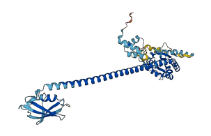
Descriptions
The autoinhibited protein was predicted that may have potential autoinhibitory elements via cis-regPred.
Autoinhibitory domains (AIDs)
Target domain |
|
Relief mechanism |
|
Assay |
cis-regPred |
Accessory elements
No accessory elements
Autoinhibited structure

Activated structure
1 structures for Q3TDN2
| Entry ID | Method | Resolution | Chain | Position | Source |
|---|---|---|---|---|---|
| AF-Q3TDN2-F1 | Predicted | AlphaFoldDB |
30 variants for Q3TDN2
| Variant ID(s) | Position | Change | Description | Diseaes Association | Provenance |
|---|---|---|---|---|---|
| rs3389284339 | 36 | L>M | No | EVA | |
| rs3389280413 | 47 | V>I | No | EVA | |
| rs3389284314 | 72 | N>S | No | EVA | |
| rs3389285776 | 76 | H>Q | No | EVA | |
| rs3389285775 | 87 | Q>* | No | EVA | |
| rs3413069134 | 119 | R>C | No | EVA | |
| rs3389283237 | 121 | I>L | No | EVA | |
| rs3389241536 | 135 | D>V | No | EVA | |
| rs3389283304 | 139 | F>I | No | EVA | |
| rs3389277024 | 146 | K>T | No | EVA | |
| rs3389292361 | 147 | Y>* | No | EVA | |
| rs3389283230 | 162 | A>T | No | EVA | |
| rs3389286338 | 167 | K>R | No | EVA | |
| rs3389286381 | 199 | I>F | No | EVA | |
| rs3404202816 | 209 | W>* | No | EVA | |
| rs3389286356 | 209 | W>R | No | EVA | |
| rs3389285765 | 224 | A>G | No | EVA | |
| rs3389285746 | 239 | K>E | No | EVA | |
| rs3389241535 | 241 | R>Q | No | EVA | |
| rs3389285759 | 262 | L>V | No | EVA | |
| rs3389292321 | 282 | E>K | No | EVA | |
| rs3389295211 | 369 | P>R | No | EVA | |
| rs3389267969 | 379 | H>L | No | EVA | |
| rs3389252604 | 379 | H>N | No | EVA | |
| rs3389292355 | 394 | L>F | No | EVA | |
| rs3389213676 | 409 | R>I | No | EVA | |
| rs3389277084 | 413 | P>T | No | EVA | |
| rs3389283269 | 420 | W>C | No | EVA | |
| rs3389295191 | 426 | L>V | No | EVA | |
| rs3404208284 | 443 | T>I | No | EVA |
No associated diseases with Q3TDN2
3 GO annotations of cellular component
| Name | Definition |
|---|---|
| endoplasmic reticulum | The irregular network of unit membranes, visible only by electron microscopy, that occurs in the cytoplasm of many eukaryotic cells. The membranes form a complex meshwork of tubular channels, which are often expanded into slitlike cavities called cisternae. The ER takes two forms, rough (or granular), with ribosomes adhering to the outer surface, and smooth (with no ribosomes attached). |
| lipid droplet | An intracellular non-membrane-bounded organelle comprising a matrix of coalesced lipids surrounded by a phospholipid monolayer. May include associated proteins. |
| VCP-NPL4-UFD1 AAA ATPase complex | A multiprotein ATPase complex required for the efficient dislocation of ER-lumenal degradation substrates, and their subsequent proteolysis by the proteasome. In budding yeast, this complex includes Cdc48p, Npl4p and Ufd1p proteins. In mammals, this complex includes a hexamer of VCP/p97 (a cytosolic ATPase) and trimers of each of its cofactors UFD1L and NPL4 (NPLOC4) (e.g. a 6:3:3 stoichiometry). |
4 GO annotations of molecular function
| Name | Definition |
|---|---|
| lipase binding | Binding to a lipase. |
| lipase inhibitor activity | Binds to and stops, prevents or reduces the activity of a lipase, an enzyme that catalyzes of the hydrolysis of a lipid. |
| ubiquitin binding | Binding to ubiquitin, a protein that when covalently bound to other cellular proteins marks them for proteolytic degradation. |
| ubiquitin protein ligase binding | Binding to a ubiquitin protein ligase enzyme, any of the E3 proteins. |
5 GO annotations of biological process
| Name | Definition |
|---|---|
| lipid droplet organization | A process that is carried out at the cellular level which results in the assembly, arrangement of constituent parts, or disassembly of a lipid particle. |
| proteasome-mediated ubiquitin-dependent protein catabolic process | The chemical reactions and pathways resulting in the breakdown of a protein or peptide by hydrolysis of its peptide bonds, initiated by the covalent attachment of ubiquitin, and mediated by the proteasome. |
| response to unfolded protein | Any process that results in a change in state or activity of a cell or an organism (in terms of movement, secretion, enzyme production, gene expression, etc.) as a result of an unfolded protein stimulus. |
| retrograde protein transport, ER to cytosol | The directed movement of unfolded or misfolded proteins from the endoplasmic reticulum to the cytosol through the translocon. |
| ubiquitin-dependent ERAD pathway | The series of steps necessary to target endoplasmic reticulum (ER)-resident proteins for degradation by the cytoplasmic proteasome. Begins with recognition of the ER-resident protein, includes retrotranslocation (dislocation) of the protein from the ER to the cytosol, protein ubiquitination necessary for correct substrate transfer, transport of the protein to the proteasome, and ends with degradation of the protein by the cytoplasmic proteasome. |
3 homologous proteins in AiPD
| UniProt AC | Gene Name | Protein Name | Species | Evidence Code |
|---|---|---|---|---|
| Q04228 | UBX2 | UBX domain-containing protein 2 | Saccharomyces cerevisiae (strain ATCC 204508 / S288c) (Baker's yeast) | PR |
| Q12229 | UBX3 | UBX domain-containing protein 3 | Saccharomyces cerevisiae (strain ATCC 204508 / S288c) (Baker's yeast) | PR |
| Q96CS3 | FAF2 | FAS-associated factor 2 | Homo sapiens (Human) | PR |
| 10 | 20 | 30 | 40 | 50 | 60 |
| MAAPEEQDLT | QEQTEKLLQF | QDLTGIESME | QCRLALEQHN | WNMEAAVQDR | LNEQEGVPSV |
| 70 | 80 | 90 | 100 | 110 | 120 |
| FNPPPARPLQ | VNTADHRIYS | YVVSRPQPRG | LLGWGYYLIM | LPFRFTYYTI | LDIFRFALRF |
| 130 | 140 | 150 | 160 | 170 | 180 |
| IRPDPRSRVT | DPVGDIVSFM | HSFEEKYGRA | HPVFYQGTYS | QALNDAKREL | RFLLVYLHGD |
| 190 | 200 | 210 | 220 | 230 | 240 |
| DHQDSDEFCR | NALCAPEVIS | LINSRMLFWA | CSTNKPEGYR | VSQALRENTY | PFLAMIMLKD |
| 250 | 260 | 270 | 280 | 290 | 300 |
| RRMTVVGRLE | GLIQPDDLIN | QLTFIMDANQ | TYLVSERLER | EERNQTQVLR | QQQDEAYLAS |
| 310 | 320 | 330 | 340 | 350 | 360 |
| LRADQEKERK | KREEKERKRR | KEEEVQQQKL | AEERRRQNLQ | EEKERKLECL | PPEPSPDDPE |
| 370 | 380 | 390 | 400 | 410 | 420 |
| SVKIIFKLPN | DSRVERRFHF | SQSLTVIHDF | LFSLKESPEK | FQIEANFPRR | VLPCVPSEEW |
| 430 | 440 | ||||
| PNPPTLQEAG | LSHTEVLFVQ | DLTDE |