Descriptions
The autoinhibited protein was predicted that may have potential autoinhibitory elements via cis-regPred.
Autoinhibitory domains (AIDs)
Target domain |
|
Relief mechanism |
|
Assay |
cis-regPred |
Accessory elements
No accessory elements
Autoinhibited structure

Activated structure
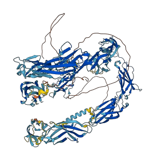
1 structures for P11627
| Entry ID | Method | Resolution | Chain | Position | Source |
|---|---|---|---|---|---|
| AF-P11627-F1 | Predicted | AlphaFoldDB |
No variants for P11627
| Variant ID(s) | Position | Change | Description | Diseaes Association | Provenance |
|---|---|---|---|---|---|
| No variants for P11627 | |||||
No associated diseases with P11627
25 regional properties for P11627
| Type | Name | Position | InterPro Accession |
|---|---|---|---|
| domain | Immunoglobulin subtype 2 | 48 - 120 | IPR003598-1 |
| domain | Immunoglobulin subtype 2 | 254 - 318 | IPR003598-2 |
| domain | Immunoglobulin subtype 2 | 344 - 410 | IPR003598-3 |
| domain | Immunoglobulin subtype 2 | 438 - 503 | IPR003598-4 |
| domain | Immunoglobulin subtype 2 | 529 - 597 | IPR003598-5 |
| domain | Immunoglobulin subtype | 42 - 132 | IPR003599-1 |
| domain | Immunoglobulin subtype | 142 - 229 | IPR003599-2 |
| domain | Immunoglobulin subtype | 248 - 329 | IPR003599-3 |
| domain | Immunoglobulin subtype | 338 - 421 | IPR003599-4 |
| domain | Immunoglobulin subtype | 432 - 514 | IPR003599-5 |
| domain | Immunoglobulin subtype | 523 - 608 | IPR003599-6 |
| domain | Fibronectin type III | 611 - 711 | IPR003961-1 |
| domain | Fibronectin type III | 714 - 809 | IPR003961-2 |
| domain | Fibronectin type III | 811 - 916 | IPR003961-3 |
| domain | Fibronectin type III | 917 - 1015 | IPR003961-4 |
| domain | Immunoglobulin-like domain | 35 - 130 | IPR007110-1 |
| domain | Immunoglobulin-like domain | 138 - 225 | IPR007110-2 |
| domain | Immunoglobulin-like domain | 239 - 327 | IPR007110-3 |
| domain | Immunoglobulin-like domain | 332 - 419 | IPR007110-4 |
| domain | Immunoglobulin-like domain | 424 - 506 | IPR007110-5 |
| domain | Immunoglobulin-like domain | 517 - 600 | IPR007110-6 |
| domain | Immunoglobulin I-set | 247 - 328 | IPR013098-1 |
| domain | Immunoglobulin I-set | 432 - 513 | IPR013098-2 |
| domain | Immunoglobulin I-set | 518 - 607 | IPR013098-3 |
| domain | Neurofascin/L1/NrCAM, C-terminal domain | 1147 - 1236 | IPR026966 |
Functions
15 GO annotations of cellular component
| Name | Definition |
|---|---|
| axon | The long process of a neuron that conducts nerve impulses, usually away from the cell body to the terminals and varicosities, which are sites of storage and release of neurotransmitter. |
| axonal growth cone | The migrating motile tip of a growing nerve cell axon. |
| cell surface | The external part of the cell wall and/or plasma membrane. |
| dendrite | A neuron projection that has a short, tapering, morphology. Dendrites receive and integrate signals from other neurons or from sensory stimuli, and conduct nerve impulses towards the axon or the cell body. In most neurons, the impulse is conveyed from dendrites to axon via the cell body, but in some types of unipolar neuron, the impulse does not travel via the cell body. |
| dendritic growth cone | The migrating motile tip of a growing nerve cell dendrite. |
| endosome | A vacuole to which materials ingested by endocytosis are delivered. |
| external side of plasma membrane | The leaflet of the plasma membrane that faces away from the cytoplasm and any proteins embedded or anchored in it or attached to its surface. |
| integral component of membrane | The component of a membrane consisting of the gene products and protein complexes having at least some part of their peptide sequence embedded in the hydrophobic region of the membrane. |
| membrane | A lipid bilayer along with all the proteins and protein complexes embedded in it an attached to it. |
| membrane raft | Any of the small (10-200 nm), heterogeneous, highly dynamic, sterol- and sphingolipid-enriched membrane domains that compartmentalize cellular processes. Small rafts can sometimes be stabilized to form larger platforms through protein-protein and protein-lipid interactions. |
| neuronal cell body | The portion of a neuron that includes the nucleus, but excludes cell projections such as axons and dendrites. |
| plasma membrane | The membrane surrounding a cell that separates the cell from its external environment. It consists of a phospholipid bilayer and associated proteins. |
| presynaptic membrane | A specialized area of membrane of the axon terminal that faces the plasma membrane of the neuron or muscle fiber with which the axon terminal establishes a synaptic junction; many synaptic junctions exhibit structural presynaptic characteristics, such as conical, electron-dense internal protrusions, that distinguish it from the remainder of the axon plasma membrane. |
| Schaffer collateral - CA1 synapse | A synapse between the Schaffer collateral axon of a CA3 pyramidal cell and a CA1 pyramidal cell. |
| terminal bouton | Terminal inflated portion of the axon, containing the specialized apparatus necessary to release neurotransmitters. The axon terminus is considered to be the whole region of thickening and the terminal bouton is a specialized region of it. |
7 GO annotations of molecular function
| Name | Definition |
|---|---|
| axon guidance receptor activity | Combining with an extracellular messenger and transmitting the signal from one side of the membrane to the other to results in a change in cellular activity involved in axon guidance. |
| identical protein binding | Binding to an identical protein or proteins. |
| integrin binding | Binding to an integrin. |
| PDZ domain binding | Binding to a PDZ domain of a protein, a domain found in diverse signaling proteins. |
| protein domain specific binding | Binding to a specific domain of a protein. |
| protein self-association | Binding to a domain within the same polypeptide. |
| sialic acid binding | Binding to a sialic acid, a N- or O- substituted derivative of neuraminic acid, a nine carbon monosaccharide. Sialic acids often occur in polysaccharides, glycoproteins, and glycolipids in animals and bacteria. |
20 GO annotations of biological process
| Name | Definition |
|---|---|
| axon development | The progression of an axon over time. Covers axonogenesis (de novo generation of an axon) and axon regeneration (regrowth), as well as processes pertaining to the progression of the axon over time (fasciculation and defasciculation). |
| axon guidance | The chemotaxis process that directs the migration of an axon growth cone to a specific target site in response to a combination of attractive and repulsive cues. |
| axonal fasciculation | The collection of axons into a bundle of rods, known as a fascicle. |
| calcium-mediated signaling | Any intracellular signal transduction in which the signal is passed on within the cell via calcium ions. |
| cell migration | The controlled self-propelled movement of a cell from one site to a destination guided by molecular cues. Cell migration is a central process in the development and maintenance of multicellular organisms. |
| cell surface receptor signaling pathway | The series of molecular signals initiated by activation of a receptor on the surface of a cell. The pathway begins with binding of an extracellular ligand to a cell surface receptor, or for receptors that signal in the absence of a ligand, by ligand-withdrawal or the activity of a constitutively active receptor. The pathway ends with regulation of a downstream cellular process, e.g. transcription. |
| cell-cell adhesion mediated by integrin | The attachment of one cell to another cell via an integrin, a heterodimeric adhesion receptor formed by the non-covalent association of particular alpha and beta subunits. |
| cell-matrix adhesion | The binding of a cell to the extracellular matrix via adhesion molecules. |
| heterophilic cell-cell adhesion via plasma membrane cell adhesion molecules | The attachment of an adhesion molecule in one cell to a nonidentical adhesion molecule in an adjacent cell. |
| homophilic cell adhesion via plasma membrane adhesion molecules | The attachment of a plasma membrane adhesion molecule in one cell to an identical molecule in an adjacent cell. |
| homotypic cell-cell adhesion | The attachment of a cell to a second cell of the identical type via adhesion molecules. |
| leukocyte cell-cell adhesion | The attachment of a leukocyte to another cell via adhesion molecules. |
| modulation of chemical synaptic transmission | Any process that modulates the frequency or amplitude of synaptic transmission, the process of communication from a neuron to a target (neuron, muscle, or secretory cell) across a synapse. Amplitude, in this case, refers to the change in postsynaptic membrane potential due to a single instance of synaptic transmission. |
| neuron projection development | The process whose specific outcome is the progression of a neuron projection over time, from its formation to the mature structure. A neuron projection is any process extending from a neural cell, such as axons or dendrites (collectively called neurites). |
| positive regulation of axon extension | Any process that activates or increases the frequency, rate or extent of axon extension. |
| positive regulation of calcium-mediated signaling | Any process that activates or increases the frequency, rate or extent of calcium-mediated signaling. |
| positive regulation of cell-cell adhesion | Any process that activates or increases the rate or extent of cell adhesion to another cell. |
| positive regulation of cell-substrate adhesion | Any process that increases the frequency, rate or extent of cell-substrate adhesion. Cell-substrate adhesion is the attachment of a cell to the underlying substrate via adhesion molecules. |
| regulation of ERK1 and ERK2 cascade | Any process that modulates the frequency, rate or extent of signal transduction mediated by the ERK1 and ERK2 cascade. |
| synapse organization | A process that is carried out at the cellular level which results in the assembly, arrangement of constituent parts, or disassembly of a synapse, the junction between a neuron and a target (neuron, muscle, or secretory cell). |
5 homologous proteins in AiPD
| UniProt AC | Gene Name | Protein Name | Species | Evidence Code |
|---|---|---|---|---|
| P32004 | L1CAM | Neural cell adhesion molecule L1 | Homo sapiens (Human) | PR |
| Q810U3 | Nfasc | Neurofascin | Mus musculus (Mouse) | PR |
| Q62845 | Cntn4 | Contactin-4 | Rattus norvegicus (Rat) | PR |
| Q62682 | Cntn3 | Contactin-3 | Rattus norvegicus (Rat) | PR |
| Q05695 | L1cam | Neural cell adhesion molecule L1 | Rattus norvegicus (Rat) | PR |
| 10 | 20 | 30 | 40 | 50 | 60 |
| MVVMLRYVWP | LLLCSPCLLI | QIPDEYKGHH | VLEPPVITEQ | SPRRLVVFPT | DDISLKCEAR |
| 70 | 80 | 90 | 100 | 110 | 120 |
| GRPQVEFRWT | KDGIHFKPKE | ELGVVVHEAP | YSGSFTIEGN | NSFAQRFQGI | YRCYASNKLG |
| 130 | 140 | 150 | 160 | 170 | 180 |
| TAMSHEIQLV | AEGAPKWPKE | TVKPVEVEEG | ESVVLPCNPP | PSAAPPRIYW | MNSKIFDIKQ |
| 190 | 200 | 210 | 220 | 230 | 240 |
| DERVSMGQNG | DLYFANVLTS | DNHSDYICNA | HFPGTRTIIQ | KEPIDLRVKP | TNSMIDRKPR |
| 250 | 260 | 270 | 280 | 290 | 300 |
| LLFPTNSSSR | LVALQGQSLI | LECIAEGFPT | PTIKWLHPSD | PMPTDRVIYQ | NHNKTLQLLN |
| 310 | 320 | 330 | 340 | 350 | 360 |
| VGEEDDGEYT | CLAENSLGSA | RHAYYVTVEA | APYWLQKPQS | HLYGPGETAR | LDCQVQGRPQ |
| 370 | 380 | 390 | 400 | 410 | 420 |
| PEITWRINGM | SMETVNKDQK | YRIEQGSLIL | SNVQPTDTMV | TQCEARNQHG | LLLANAYIYV |
| 430 | 440 | 450 | 460 | 470 | 480 |
| VQLPARILTK | DNQTYMAVEG | STAYLLCKAF | GAPVPSVQWL | DEEGTTVLQD | ERFFPYANGT |
| 490 | 500 | 510 | 520 | 530 | 540 |
| LSIRDLQAND | TGRYFCQAAN | DQNNVTILAN | LQVKEATQIT | QGPRSAIEKK | GARVTFTCQA |
| 550 | 560 | 570 | 580 | 590 | 600 |
| SFDPSLQASI | TWRGDGRDLQ | ERGDSDKYFI | EDGKLVIQSL | DYSDQGNYSC | VASTELDEVE |
| 610 | 620 | 630 | 640 | 650 | 660 |
| SRAQLLVVGS | PGPVPHLELS | DRHLLKQSQV | HLSWSPAEDH | NSPIEKYDIE | FEDKEMAPEK |
| 670 | 680 | 690 | 700 | 710 | 720 |
| WFSLGKVPGN | QTSTTLKLSP | YVHYTFRVTA | INKYGPGEPS | PVSESVVTPE | AAPEKNPVDV |
| 730 | 740 | 750 | 760 | 770 | 780 |
| RGEGNETNNM | VITWKPLRWM | DWNAPQIQYR | VQWRPQGKQE | TWRKQTVSDP | FLVVSNTSTF |
| 790 | 800 | 810 | 820 | 830 | 840 |
| VPYEIKVQAV | NNQGKGPEPQ | VTIGYSGEDY | PQVSPELEDI | TIFNSSTVLV | RWRPVDLAQV |
| 850 | 860 | 870 | 880 | 890 | 900 |
| KGHLKGYNVT | YWWKGSQRKH | SKRHIHKSHI | VVPANTTSAI | LSGLRPYSSY | HVEVQAFNGR |
| 910 | 920 | 930 | 940 | 950 | 960 |
| GLGPASEWTF | STPEGVPGHP | EALHLECQSD | TSLLLHWQPP | LSHNGVLTGY | LLSYHPVEGE |
| 970 | 980 | 990 | 1000 | 1010 | 1020 |
| SKEQLFFNLS | DPELRTHNLT | NLNPDLQYRF | QLQATTQQGG | PGEAIVREGG | TMALFGKPDF |
| 1030 | 1040 | 1050 | 1060 | 1070 | 1080 |
| GNISATAGEN | YSVVSWVPRK | GQCNFRFHIL | FKALPEGKVS | PDHQPQPQYV | SYNQSSYTQW |
| 1090 | 1100 | 1110 | 1120 | 1130 | 1140 |
| NLQPDTKYEI | HLIKEKVLLH | HLDVKTNGTG | PVRVSTTGSF | ASEGWFIAFV | SAIILLLLIL |
| 1150 | 1160 | 1170 | 1180 | 1190 | 1200 |
| LILCFIKRSK | GGKYSVKDKE | DTQVDSEARP | MKDETFGEYR | SLESDNEEKA | FGSSQPSLNG |
| 1210 | 1220 | 1230 | 1240 | 1250 | |
| DIKPLGSDDS | LADYGGSVDV | QFNEDGSFIG | QYSGKKEKEA | AGGNDSSGAT | SPINPAVALE |