Q3T040
Gene name |
MRPS7 |
Protein name |
28S ribosomal protein S7, mitochondrial |
Names |
MRP-S7, S7mt |
Species |
Bos taurus (Bovine) |
KEGG Pathway |
bta:509115 |
EC number |
|
Protein Class |
|
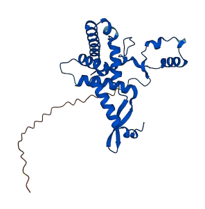
Descriptions
The autoinhibited protein was predicted that may have potential autoinhibitory elements via cis-regPred.
Autoinhibitory domains (AIDs)
Target domain |
|
Relief mechanism |
|
Assay |
cis-regPred |
Accessory elements
No accessory elements
Autoinhibited structure

Activated structure
4 structures for Q3T040
| Entry ID | Method | Resolution | Chain | Position | Source |
|---|---|---|---|---|---|
| 3JD5 | EM | 700 A | G | 1-242 | PDB |
| 6NEQ | EM | 332 A | G | 1-242 | PDB |
| 6NF8 | EM | 348 A | G | 1-242 | PDB |
| AF-Q3T040-F1 | Predicted | AlphaFoldDB |
48 variants for Q3T040
| Variant ID(s) | Position | Change | Description | Diseaes Association | Provenance |
|---|---|---|---|---|---|
| rs476339003 | 4 | P>S | No | EVA | |
| rs524259022 | 5 | T>M | No | EVA | |
| rs433345818 | 5 | T>P | No | EVA | |
| rs519036131 | 6 | A>V | No | EVA | |
| rs434288469 | 9 | S>A | No | EVA | |
| rs468523196 | 15 | L>S | No | EVA | |
| rs469497226 | 20 | R>P | No | EVA | |
| rs446136949 | 21 | I>V | No | EVA | |
| rs477773169 | 23 | V>A | No | EVA | |
| rs476099607 | 30 | T>S | No | EVA | |
| rs459132872 | 34 | W>G | No | EVA | |
| rs438740607 | 71 | T>P | No | EVA | |
| rs207718097 | 79 | A>P | No | EVA | |
| rs437833195 | 132 | E>V | No | EVA | |
| rs477383961 | 137 | V>I | No | EVA | |
| rs453212020 | 170 | V>G | No | EVA | |
| rs435973320 | 175 | A>T | No | EVA | |
| rs467407730 | 177 | R>P | No | EVA | |
| rs717568439 | 178 | R>H | No | EVA | |
| rs456947717 | 180 | R>P | No | EVA | |
| rs436860868 | 181 | F>C | No | EVA | |
| rs465356182 | 185 | K>E | No | EVA | |
| rs465356182 | 185 | K>Q | No | EVA | |
| rs445390072 | 186 | W>G | No | EVA | |
| rs445390072 | 186 | W>R | No | EVA | |
| rs449598228 | 187 | M>I | No | EVA | |
| rs479864211 | 187 | M>L | No | EVA | |
| rs466402897 | 187 | M>R | No | EVA | |
| rs480638532 | 188 | I>T | No | EVA | |
| rs443704630 | 190 | E>G | No | EVA | |
| rs478291931 | 191 | C>G | No | EVA | |
| rs478291931 | 191 | C>R | No | EVA | |
| rs458278788 | 194 | K>E | No | EVA | |
| rs438599122 | 194 | K>R | No | EVA | |
| rs453097606 | 198 | R>G | No | EVA | |
| rs442735029 | 204 | K>E | No | EVA | |
| rs473752712 | 205 | L>M | No | EVA | |
| rs456928011 | 205 | L>R | No | EVA | |
| rs464890509 | 211 | E>G | No | EVA | |
| rs451324438 | 216 | R>C | No | EVA | |
| rs434935878 | 221 | K>R | No | EVA | |
| rs449470031 | 224 | H>Y | No | EVA | |
| rs435898552 | 229 | M>K | No | EVA | |
| rs470512949 | 231 | E>D | No | EVA | |
| rs450102572 | 232 | A>D | No | EVA | |
| rs458218819 | 234 | R>S | No | EVA | |
| rs459550341 | 237 | A>D | No | EVA | |
| rs442650660 | 242 | W>R | No | EVA |
No associated diseases with Q3T040
1 regional properties for Q3T040
| Type | Name | Position | InterPro Accession |
|---|---|---|---|
| domain | Ribosomal protein S7 domain | 79 - 234 | IPR023798 |
3 GO annotations of cellular component
| Name | Definition |
|---|---|
| mitochondrial inner membrane | The inner, i.e. lumen-facing, lipid bilayer of the mitochondrial envelope. It is highly folded to form cristae. |
| mitochondrial small ribosomal subunit | The smaller of the two subunits of a mitochondrial ribosome. |
| ribosome | An intracellular organelle, about 200 A in diameter, consisting of RNA and protein. It is the site of protein biosynthesis resulting from translation of messenger RNA (mRNA). It consists of two subunits, one large and one small, each containing only protein and RNA. Both the ribosome and its subunits are characterized by their sedimentation coefficients, expressed in Svedberg units (symbol: S). Hence, the prokaryotic ribosome (70S) comprises a large (50S) subunit and a small (30S) subunit, while the eukaryotic ribosome (80S) comprises a large (60S) subunit and a small (40S) subunit. Two sites on the ribosomal large subunit are involved in translation, namely the aminoacyl site (A site) and peptidyl site (P site). Ribosomes from prokaryotes, eukaryotes, mitochondria, and chloroplasts have characteristically distinct ribosomal proteins. |
3 GO annotations of molecular function
| Name | Definition |
|---|---|
| mRNA binding | Binding to messenger RNA (mRNA), an intermediate molecule between DNA and protein. mRNA includes UTR and coding sequences, but does not contain introns. |
| rRNA binding | Binding to a ribosomal RNA. |
| structural constituent of ribosome | The action of a molecule that contributes to the structural integrity of the ribosome. |
3 GO annotations of biological process
| Name | Definition |
|---|---|
| mitochondrial translation | The chemical reactions and pathways resulting in the formation of a protein in a mitochondrion. This is a ribosome-mediated process in which the information in messenger RNA (mRNA) is used to specify the sequence of amino acids in the protein; the mitochondrion has its own ribosomes and transfer RNAs, and uses a genetic code that differs from the nuclear code. |
| ribosomal small subunit assembly | The aggregation, arrangement and bonding together of constituent RNAs and proteins to form the small ribosomal subunit. |
| translation | The cellular metabolic process in which a protein is formed, using the sequence of a mature mRNA or circRNA molecule to specify the sequence of amino acids in a polypeptide chain. Translation is mediated by the ribosome, and begins with the formation of a ternary complex between aminoacylated initiator methionine tRNA, GTP, and initiation factor 2, which subsequently associates with the small subunit of the ribosome and an mRNA or circRNA. Translation ends with the release of a polypeptide chain from the ribosome. |
8 homologous proteins in AiPD
| UniProt AC | Gene Name | Protein Name | Species | Evidence Code |
|---|---|---|---|---|
| P47150 | RSM7 | 37S ribosomal protein S7, mitochondrial | Saccharomyces cerevisiae (strain ATCC 204508 / S288c) (Baker's yeast) | PR |
| Q24186 | RpS5a | 40S ribosomal protein S5a | Drosophila melanogaster (Fruit fly) | PR |
| Q9VFE4 | RpS5b | 40S ribosomal protein S5b | Drosophila melanogaster (Fruit fly) | PR |
| Q9Y2R9 | MRPS7 | 28S ribosomal protein S7, mitochondrial | Homo sapiens (Human) | PR |
| Q80X85 | Mrps7 | 28S ribosomal protein S7, mitochondrial | Mus musculus (Mouse) | PR |
| Q5I0K8 | Mrps7 | 28S ribosomal protein S7, mitochondrial | Rattus norvegicus (Rat) | PR |
| P49041 | rps-5 | 40S ribosomal protein S5 | Caenorhabditis elegans | PR |
| Q498Z6 | mrps7 | 28S ribosomal protein S7, mitochondrial | Danio rerio (Zebrafish) (Brachydanio rerio) | PR |
| 10 | 20 | 30 | 40 | 50 | 60 |
| MAAPTAKVSR | GWSGLALGVR | IAVLRLPGLT | QVRWSRYGPE | YQDPQIDKEY | YRKPLAQLTE |
| 70 | 80 | 90 | 100 | 110 | 120 |
| EETYERELRK | TQVIKAAPAT | KTSSVFEDPV | ISKFTNMMMK | GGNKILARSL | MTQTLEAVKR |
| 130 | 140 | 150 | 160 | 170 | 180 |
| KQFEKYHAAS | AEEQATVERN | PYTIFHQALK | NCEPVIGLVP | ILKGGHFYQV | PVPLAERRRR |
| 190 | 200 | 210 | 220 | 230 | 240 |
| FLAMKWMITE | CREKKPRRML | MPEKLSQELL | EAFCNRGPVI | KRKHDMHKMA | EANRALAHYR |
| WW |