P32610
Gene name |
VMA8 |
Protein name |
V-type proton ATPase subunit D |
Names |
V-ATPase subunit D, Vacuolar proton pump subunit D |
Species |
Saccharomyces cerevisiae (strain ATCC 204508 / S288c) (Baker's yeast) |
KEGG Pathway |
sce:YEL051W |
EC number |
|
Protein Class |
|
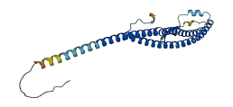
Descriptions
The autoinhibited protein was predicted that may have potential autoinhibitory elements via cis-regPred.
Autoinhibitory domains (AIDs)
Target domain |
|
Relief mechanism |
|
Assay |
cis-regPred |
Accessory elements
No accessory elements
Autoinhibited structure

Activated structure
22 structures for P32610
| Entry ID | Method | Resolution | Chain | Position | Source |
|---|---|---|---|---|---|
| 3J9T | EM | 690 A | M | 1-256 | PDB |
| 3J9U | EM | 760 A | M | 1-256 | PDB |
| 3J9V | EM | 830 A | M | 1-256 | PDB |
| 4RND | X-ray | 318 A | A/C | 1-256 | PDB |
| 5BW9 | X-ray | 700 A | G/g | 1-256 | PDB |
| 5D80 | X-ray | 620 A | G/g | 1-256 | PDB |
| 5VOX | EM | 680 A | M | 1-256 | PDB |
| 5VOY | EM | 790 A | M | 1-256 | PDB |
| 5VOZ | EM | 760 A | M | 1-256 | PDB |
| 6O7V | EM | 660 A | M | 1-256 | PDB |
| 6O7W | EM | 700 A | M | 1-256 | PDB |
| 6O7X | EM | 870 A | M | 1-256 | PDB |
| 7FDA | EM | 420 A | M | 1-256 | PDB |
| 7FDB | EM | 480 A | M | 1-256 | PDB |
| 7FDC | EM | 660 A | M | 1-256 | PDB |
| 7FDE | EM | 380 A | M | 1-256 | PDB |
| 7TMM | EM | 350 A | M | 1-256 | PDB |
| 7TMO | EM | 330 A | M | 1-256 | PDB |
| 7TMP | EM | 330 A | M | 1-256 | PDB |
| 7TMQ | EM | 330 A | M | 1-256 | PDB |
| 7TMR | EM | 350 A | M | 1-256 | PDB |
| AF-P32610-F1 | Predicted | AlphaFoldDB |
1 variants for P32610
| Variant ID(s) | Position | Change | Description | Diseaes Association | Provenance |
|---|---|---|---|---|---|
| s05-59099 | 241 | P>R | No | SGRP |
No associated diseases with P32610
No regional properties for P32610
| Type | Name | Position | InterPro Accession |
|---|---|---|---|
| No domain, repeats, and functional sites for P32610 | |||
8 GO annotations of cellular component
| Name | Definition |
|---|---|
| endosome membrane | The lipid bilayer surrounding an endosome. |
| fungal-type vacuole membrane | The lipid bilayer surrounding a vacuole, the shape of which correlates with cell cycle phase. The membrane separates its contents from the cytoplasm of the cell. An example of this structure is found in Saccharomyces cerevisiae. |
| Golgi membrane | The lipid bilayer surrounding any of the compartments of the Golgi apparatus. |
| membrane | A lipid bilayer along with all the proteins and protein complexes embedded in it an attached to it. |
| proton-transporting V-type ATPase complex | A proton-transporting two-sector ATPase complex that couples ATP hydrolysis to the transport of protons across a concentration gradient. The resulting transmembrane electrochemical potential of H+ is used to drive a variety of (i) secondary active transport systems via H+-dependent symporters and antiporters and (ii) channel-mediated transport systems. The complex comprises a membrane sector (V0) that carries out proton transport and a cytoplasmic compartment sector (V1) that catalyzes ATP hydrolysis. V-type ATPases are found in the membranes of organelles such as vacuoles, endosomes, and lysosomes, and in the plasma membrane. |
| vacuolar membrane | The lipid bilayer surrounding the vacuole and separating its contents from the cytoplasm of the cell. |
| vacuolar proton-transporting V-type ATPase complex | A proton-transporting two-sector ATPase complex found in the vacuolar membrane, where it acts as a proton pump to mediate acidification of the vacuolar lumen. |
| vacuolar proton-transporting V-type ATPase, V1 domain | The V1 domain of a proton-transporting V-type ATPase found in the vacuolar membrane. |
1 GO annotations of molecular function
| Name | Definition |
|---|---|
| proton-transporting ATPase activity, rotational mechanism | Enables the transfer of protons from one side of a membrane to the other according to the reaction: ATP + H2O + H+(in) = ADP + phosphate + H+(out), by a rotational mechanism. |
4 GO annotations of biological process
| Name | Definition |
|---|---|
| endosomal lumen acidification | Any process that reduces the pH of the endosomal lumen, measured by the concentration of the hydrogen ion. |
| Golgi lumen acidification | Any process that reduces the pH of the Golgi lumen, measured by the concentration of the hydrogen ion. |
| proton transmembrane transport | The directed movement of a proton across a membrane. |
| vacuolar acidification | Any process that reduces the pH of the vacuole, measured by the concentration of the hydrogen ion. |
3 homologous proteins in AiPD
| 10 | 20 | 30 | 40 | 50 | 60 |
| MSGNREQVFP | TRMTLGLMKT | KLKGANQGYS | LLKRKSEALT | KRFRDITKRI | DDAKQKMGRV |
| 70 | 80 | 90 | 100 | 110 | 120 |
| MQTAAFSLAE | VSYATGENIG | YQVQESVSTA | RFKVRARQEN | VSGVYLSQFE | SYIDPEINDF |
| 130 | 140 | 150 | 160 | 170 | 180 |
| RLTGLGRGGQ | QVQRAKEIYS | RAVETLVELA | SLQTAFIILD | EVIKVTNRRV | NAIEHVIIPR |
| 190 | 200 | 210 | 220 | 230 | 240 |
| TENTIAYINS | ELDELDREEF | YRLKKVQEKK | QNETAKLDAE | MKLKRDRAEQ | DASEVAADEE |
| 250 | |||||
| PQGETLVADQ | EDDVIF |