Q9JIF9
Gene name |
Myot (Myo, Ttid) |
Protein name |
Myotilin |
Names |
Myofibrillar titin-like Ig domains protein, Titin immunoglobulin domain protein |
Species |
Mus musculus (Mouse) |
KEGG Pathway |
mmu:58916 |
EC number |
|
Protein Class |
|
Descriptions
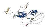
The autoinhibited protein was predicted that may have potential autoinhibitory elements via cis-regPred.
Autoinhibitory domains (AIDs)
Target domain |
|
Relief mechanism |
|
Assay |
cis-regPred |
Accessory elements
No accessory elements
Autoinhibited structure

Activated structure
1 structures for Q9JIF9
| Entry ID | Method | Resolution | Chain | Position | Source |
|---|---|---|---|---|---|
| AF-Q9JIF9-F1 | Predicted | AlphaFoldDB |
26 variants for Q9JIF9
| Variant ID(s) | Position | Change | Description | Diseaes Association | Provenance |
|---|---|---|---|---|---|
| rs3389459105 | 7 | P>S | No | EVA | |
| rs218289012 | 13 | P>S | No | EVA | |
| rs233178503 | 30 | G>D | No | EVA | |
| rs262423219 | 30 | G>S | No | EVA | |
| rs266229693 | 72 | P>S | No | EVA | |
| rs3389513585 | 87 | T>I | No | EVA | |
| rs3389510011 | 157 | I>N | No | EVA | |
| rs3389494851 | 160 | L>* | No | EVA | |
| rs3389500627 | 168 | D>H | No | EVA | |
| rs229292451 | 206 | Q>H | No | EVA | |
| rs3389494083 | 244 | K>M | No | EVA | |
| rs3389479104 | 316 | A>T | No | EVA | |
| rs3389513570 | 327 | A>T | No | EVA | |
| rs3389459124 | 330 | A>P | No | EVA | |
| rs3389509990 | 331 | T>I | No | EVA | |
| rs3389468606 | 382 | R>G | No | EVA | |
| rs3389501690 | 440 | A>D | No | EVA | |
| rs30173993 | 443 | I>N | No | EVA | |
| rs3389459100 | 448 | A>S | No | EVA | |
| rs3408861768 | 453 | R>S | No | EVA | |
| rs3389500689 | 454 | V>A | No | EVA | |
| rs31937418 | 455 | R>P | No | EVA | |
| rs3408578374 | 456 | P>R | No | EVA | |
| rs3407434846 | 458 | F>C | No | EVA | |
| rs3408427521 | 463 | A>E | No | EVA | |
| rs3408666624 | 497 | L>S | No | EVA |
2 associated diseases with Q9JIF9
[MIM: 614464]: Joubert syndrome 15 (JBTS15)
An autosomal recessive disorder presenting with cerebellar ataxia, oculomotor apraxia, hypotonia, neonatal breathing abnormalities and psychomotor delay. Neuroradiologically, it is characterized by cerebellar vermian hypoplasia/aplasia, thickened and reoriented superior cerebellar peduncles, and an abnormally large interpeduncular fossa, giving the appearance of a molar tooth on transaxial slices (molar tooth sign). Additional variable features include retinal dystrophy, renal disease, liver fibrosis and polydactyly. {ECO:0000269|PubMed:22246503}. Note=The disease is caused by variants affecting the gene represented in this entry.
Without disease ID
- An autosomal recessive disorder presenting with cerebellar ataxia, oculomotor apraxia, hypotonia, neonatal breathing abnormalities and psychomotor delay. Neuroradiologically, it is characterized by cerebellar vermian hypoplasia/aplasia, thickened and reoriented superior cerebellar peduncles, and an abnormally large interpeduncular fossa, giving the appearance of a molar tooth on transaxial slices (molar tooth sign). Additional variable features include retinal dystrophy, renal disease, liver fibrosis and polydactyly. {ECO:0000269|PubMed:22246503}. Note=The disease is caused by variants affecting the gene represented in this entry.
11 regional properties for Q9JIF9
| Type | Name | Position | InterPro Accession |
|---|---|---|---|
| domain | Cadherin-like | 53 - 130 | IPR002126-1 |
| domain | Cadherin-like | 133 - 241 | IPR002126-2 |
| domain | Cadherin-like | 241 - 345 | IPR002126-3 |
| domain | Cadherin-like | 346 - 559 | IPR002126-4 |
| domain | Cadherin-like | 574 - 669 | IPR002126-5 |
| domain | Cadherin, N-terminal | 30 - 111 | IPR013164 |
| conserved_site | Cadherin conserved site | 229 - 239 | IPR020894-1 |
| conserved_site | Cadherin conserved site | 333 - 343 | IPR020894-2 |
| conserved_site | Cadherin conserved site | 437 - 447 | IPR020894-3 |
| conserved_site | Cadherin conserved site | 547 - 557 | IPR020894-4 |
| domain | Cadherin, cytoplasmic C-terminal domain | 684 - 766 | IPR032455 |
Functions
5 GO annotations of cellular component
| Name | Definition |
|---|---|
| axon | The long process of a neuron that conducts nerve impulses, usually away from the cell body to the terminals and varicosities, which are sites of storage and release of neurotransmitter. |
| cytoskeleton | A cellular structure that forms the internal framework of eukaryotic and prokaryotic cells. The cytoskeleton includes intermediate filaments, microfilaments, microtubules, the microtrabecular lattice, and other structures characterized by a polymeric filamentous nature and long-range order within the cell. The various elements of the cytoskeleton not only serve in the maintenance of cellular shape but also have roles in other cellular functions, including cellular movement, cell division, endocytosis, and movement of organelles. |
| plasma membrane | The membrane surrounding a cell that separates the cell from its external environment. It consists of a phospholipid bilayer and associated proteins. |
| sarcolemma | The outer membrane of a muscle cell, consisting of the plasma membrane, a covering basement membrane (about 100 nm thick and sometimes common to more than one fiber), and the associated loose network of collagen fibers. |
| Z disc | Platelike region of a muscle sarcomere to which the plus ends of actin filaments are attached. |
3 GO annotations of molecular function
| Name | Definition |
|---|---|
| actin binding | Binding to monomeric or multimeric forms of actin, including actin filaments. |
| alpha-actinin binding | Binding to alpha-actinin, one of a family of proteins that cross-link F-actin as antiparallel homodimers. Alpha-actinin has a molecular mass of 93-103 KDa; at the N-terminus there are two calponin homology domains, at the C-terminus there are two EF-hands. These two domains are connected by the rod domain. This domain is formed by triple-helical spectrin repeats. |
| cell-cell adhesion mediator activity | The binding by a cell-adhesion protein on the cell surface to an extracellular matrix component, to mediate adhesion of the cell to another cell. |
3 GO annotations of biological process
| Name | Definition |
|---|---|
| axon guidance | The chemotaxis process that directs the migration of an axon growth cone to a specific target site in response to a combination of attractive and repulsive cues. |
| dendrite self-avoidance | The process in which dendrites recognize and avoid contact with sister dendrites from the same cell. |
| homophilic cell adhesion via plasma membrane adhesion molecules | The attachment of a plasma membrane adhesion molecule in one cell to an identical molecule in an adjacent cell. |
1 homologous proteins in AiPD
| UniProt AC | Gene Name | Protein Name | Species | Evidence Code |
|---|---|---|---|---|
| Q7TPW1 | Nexn | Nexilin | Mus musculus (Mouse) | PR |
| 10 | 20 | 30 | 40 | 50 | 60 |
| MFNYERPKHF | IQPQNPCGSR | LQPPGPEVSG | FPSQTKQSSI | VIQPRQCTEQ | RFSASSTVSS |
| 70 | 80 | 90 | 100 | 110 | 120 |
| HITVSSSAYP | APQQLAGPNP | GQKVTATYNQ | SPASFLSSIL | PSQPDYCNSK | IPSTVDSNYQ |
| 130 | 140 | 150 | 160 | 170 | 180 |
| QSSVNQPVNA | MSSQAANARP | TPKTPDHEIQ | GSKEALIQDL | ERKLKCKDTL | LHNGNQRLTY |
| 190 | 200 | 210 | 220 | 230 | 240 |
| EEKMARRLLG | PQNAAAVFQA | QNSDVQDSPQ | HNPEQARLHV | PTSQVRSRSS | SRAEANDQDA |
| 250 | 260 | 270 | 280 | 290 | 300 |
| IQEKFYPPRF | IQVPENMSIE | EGRFCRMDFK | VSGLPAPDVS | WYLNGRPVQS | DELHKMIVSE |
| 310 | 320 | 330 | 340 | 350 | 360 |
| KGFHSLIFEV | VRASDAGPYA | CVARNRAGEA | TFTVQLDVLA | KEHKRAPMFI | FKPQSKKVFE |
| 370 | 380 | 390 | 400 | 410 | 420 |
| GETVKLECQI | SAIPPPKLFW | KRNNEMVQFN | TDRISLYHDN | AGRVTLLIKD | VNKKDAGWYT |
| 430 | 440 | 450 | 460 | 470 | 480 |
| VSAVNEAGVT | TCNTRLDVTA | RPIQTLPAPK | QLRVRPTFSK | YLALNGRGLD | VKQAFNPEGE |
| 490 | |||||
| FQRLAAQSGL | YESEEL |