Q8NI17
Gene name |
IL31RA (CRL3, GPL, UNQ6368/PRO21073/PRO21384) |
Protein name |
Interleukin-31 receptor subunit alpha |
Names |
IL-31 receptor subunit alpha, IL-31R subunit alpha, IL-31R-alpha, IL-31RA, Cytokine receptor-like 3, GLM-R, hGLM-R, Gp130-like monocyte receptor, Gp130-like receptor, ZcytoR17 |
Species |
Homo sapiens (Human) |
KEGG Pathway |
hsa:133396 |
EC number |
|
Protein Class |
|
Descriptions
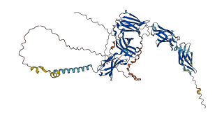
The autoinhibited protein was predicted that may have potential autoinhibitory elements via cis-regPred.
Autoinhibitory domains (AIDs)
Target domain |
|
Relief mechanism |
|
Assay |
cis-regPred |
Accessory elements
No accessory elements
Autoinhibited structure

Activated structure
1 structures for Q8NI17
| Entry ID | Method | Resolution | Chain | Position | Source |
|---|---|---|---|---|---|
| AF-Q8NI17-F1 | Predicted | AlphaFoldDB |
3 variants for Q8NI17
| Variant ID(s) | Position | Change | Description | Diseaes Association | Provenance |
|---|---|---|---|---|---|
| VAR_065809 | 489 | S>F | PLCA2 [UniProt] | Yes | UniProt |
|
VAR_030328 rs13184107 |
155 | D>N | No |
UniProt dbSNP |
|
|
rs161704 VAR_030329 |
497 | S>N | No |
UniProt dbSNP |
1 associated diseases with Q8NI17
[MIM: 613955]: Amyloidosis, primary localized cutaneous, 2 (PLCA2)
A primary amyloidosis characterized by localized cutaneous amyloid deposition. This condition usually presents with itching (especially on the lower legs) and visible changes of skin hyperpigmentation and thickening that may be exacerbated by chronic scratching and rubbing. Primary localized cutaneous amyloidosis is often divided into macular and lichen subtypes although many affected individuals often show both variants coexisting. Lichen amyloidosis characteristically presents as a pruritic eruption of grouped hyperkeratotic papules with a predilection for the shins, calves, ankles and dorsa of feet and thighs. Papules may coalesce to form hyperkeratotic plaques that can resemble lichen planus, lichen simplex or nodular prurigo. Macular amyloidosis is characterized by small pigmented macules that may merge to produce macular hyperpigmentation, sometimes with a reticulate or rippled pattern. In macular and lichen amyloidosis, amyloid is deposited in the papillary dermis in association with grouped colloid bodies, thought to represent degenerate basal keratinocytes. The amyloid deposits probably reflect a combination of degenerate keratin filaments, serum amyloid P component, and deposition of immunoglobulins. {ECO:0000269|PubMed:19690585}. Note=The disease is caused by variants affecting the gene represented in this entry.
Without disease ID
- A primary amyloidosis characterized by localized cutaneous amyloid deposition. This condition usually presents with itching (especially on the lower legs) and visible changes of skin hyperpigmentation and thickening that may be exacerbated by chronic scratching and rubbing. Primary localized cutaneous amyloidosis is often divided into macular and lichen subtypes although many affected individuals often show both variants coexisting. Lichen amyloidosis characteristically presents as a pruritic eruption of grouped hyperkeratotic papules with a predilection for the shins, calves, ankles and dorsa of feet and thighs. Papules may coalesce to form hyperkeratotic plaques that can resemble lichen planus, lichen simplex or nodular prurigo. Macular amyloidosis is characterized by small pigmented macules that may merge to produce macular hyperpigmentation, sometimes with a reticulate or rippled pattern. In macular and lichen amyloidosis, amyloid is deposited in the papillary dermis in association with grouped colloid bodies, thought to represent degenerate basal keratinocytes. The amyloid deposits probably reflect a combination of degenerate keratin filaments, serum amyloid P component, and deposition of immunoglobulins. {ECO:0000269|PubMed:19690585}. Note=The disease is caused by variants affecting the gene represented in this entry.
5 regional properties for Q8NI17
| Type | Name | Position | InterPro Accession |
|---|---|---|---|
| domain | Fibronectin type III | 22 - 122 | IPR003961-1 |
| domain | Fibronectin type III | 122 - 225 | IPR003961-2 |
| domain | Fibronectin type III | 325 - 409 | IPR003961-3 |
| domain | Fibronectin type III | 421 - 515 | IPR003961-4 |
| domain | Type I cytokine receptor, cytokine-binding domain | 27 - 116 | IPR015321 |
7 GO annotations of cellular component
| Name | Definition |
|---|---|
| anchoring junction | A cell junction that mechanically attaches a cell (and its cytoskeleton) to neighboring cells or to the extracellular matrix. |
| axon | The long process of a neuron that conducts nerve impulses, usually away from the cell body to the terminals and varicosities, which are sites of storage and release of neurotransmitter. |
| external side of plasma membrane | The leaflet of the plasma membrane that faces away from the cytoplasm and any proteins embedded or anchored in it or attached to its surface. |
| integral component of membrane | The component of a membrane consisting of the gene products and protein complexes having at least some part of their peptide sequence embedded in the hydrophobic region of the membrane. |
| plasma membrane | The membrane surrounding a cell that separates the cell from its external environment. It consists of a phospholipid bilayer and associated proteins. |
| presynaptic membrane | A specialized area of membrane of the axon terminal that faces the plasma membrane of the neuron or muscle fiber with which the axon terminal establishes a synaptic junction; many synaptic junctions exhibit structural presynaptic characteristics, such as conical, electron-dense internal protrusions, that distinguish it from the remainder of the axon plasma membrane. |
| receptor complex | Any protein complex that undergoes combination with a hormone, neurotransmitter, drug or intracellular messenger to initiate a change in cell function. |
5 GO annotations of molecular function
| Name | Definition |
|---|---|
| cytokine binding | Binding to a cytokine, any of a group of proteins that function to control the survival, growth and differentiation of tissues and cells, and which have autocrine and paracrine activity. |
| cytokine receptor activity | Combining with a cytokine and transmitting the signal from one side of the membrane to the other to initiate a change in cell activity. |
| granulocyte colony-stimulating factor binding | Binding to granulocyte colony-stimulating factor, G-CSF. |
| protein kinase binding | Binding to a protein kinase, any enzyme that catalyzes the transfer of a phosphate group, usually from ATP, to a protein substrate. |
| transcription coactivator activity | A transcription coregulator activity that activates or increases the transcription of specific gene sets via binding to a DNA-bound DNA-binding transcription factor, either on its own or as part of a complex. Coactivators often act by altering chromatin structure and modifications. For example, one class of transcription coactivators modifies chromatin structure through covalent modification of histones. A second class remodels the conformation of chromatin in an ATP-dependent fashion. A third class modulates interactions of DNA-bound DNA-binding transcription factors with other transcription coregulators. A fourth class of coactivator activity is the bridging of a DNA-binding transcription factor to the general (basal) transcription machinery. The Mediator complex, which bridges sequence-specific DNA binding transcription factors and RNA polymerase, is also a transcription coactivator. |
16 GO annotations of biological process
| Name | Definition |
|---|---|
| acute inflammatory response to antigenic stimulus | An acute inflammatory response to an antigenic stimulus. An acute inflammatory response occurs within a matter of minutes or hours, and either resolves within a few days or becomes a chronic inflammatory response. |
| cytokine-mediated signaling pathway | The series of molecular signals initiated by the binding of a cytokine to a receptor on the surface of a cell, and ending with the regulation of a downstream cellular process, e.g. transcription. |
| defense response | Reactions, triggered in response to the presence of a foreign body or the occurrence of an injury, which result in restriction of damage to the organism attacked or prevention/recovery from the infection caused by the attack. |
| defense response to other organism | Reactions triggered in response to the presence of another organism that act to protect the cell or organism from damage caused by that organism. |
| glandular epithelial cell differentiation | The process in which a relatively unspecialized cell acquires specialized features of a glandular epithelial cell. A glandular epithelial cell is a columnar/cuboidal epithelial cell found in a two dimensional sheet with a free surface exposed to the lumen of a gland. |
| homeostatic process | Any biological process involved in the maintenance of an internal steady state. |
| macrophage differentiation | The process in which a relatively unspecialized monocyte acquires the specialized features of a macrophage. |
| MAPK cascade | An intracellular protein kinase cascade containing at least a MAPK, a MAPKK and a MAP3K. The cascade can also contain an additional tiers: the upstream MAP4K. The kinases in each tier phosphorylate and activate the kinase in the downstream tier to transmit a signal within a cell. |
| monocyte differentiation | The process in which a relatively unspecialized myeloid precursor cell acquires the specialized features of a monocyte. |
| negative regulation of apoptotic process | Any process that stops, prevents, or reduces the frequency, rate or extent of cell death by apoptotic process. |
| negative regulation of macrophage activation | Any process that stops, prevents, or reduces the frequency, rate or extent of macrophage activation. |
| positive regulation of cell population proliferation | Any process that activates or increases the rate or extent of cell proliferation. |
| positive regulation of DNA-templated transcription | Any process that activates or increases the frequency, rate or extent of cellular DNA-templated transcription. |
| positive regulation of tyrosine phosphorylation of STAT protein | Any process that activates or increases the frequency, rate or extent of the introduction of a phosphate group to a tyrosine residue of a STAT (Signal Transducer and Activator of Transcription) protein. |
| receptor signaling pathway via JAK-STAT | Any process in which STAT proteins (Signal Transducers and Activators of Transcription) and JAK (Janus Activated Kinase) proteins convey a signal to trigger a change in the activity or state of a cell. The receptor signaling pathway via JAK-STAT begins with activation of a receptor and proceeeds through STAT protein activation by members of the JAK family of tyrosine kinases. STAT proteins dimerize and subsequently translocate to the nucleus. The pathway ends with regulation of target gene expression by STAT proteins. |
| transmembrane receptor protein tyrosine kinase signaling pathway | The series of molecular signals initiated by an extracellular ligand binding to a receptor on the surface of the target cell where the receptor possesses tyrosine kinase activity, and ending with the regulation of a downstream cellular process, e.g. transcription. |
1 homologous proteins in AiPD
| UniProt AC | Gene Name | Protein Name | Species | Evidence Code |
|---|---|---|---|---|
| Q8K5B1 | Il31ra | Interleukin-31 receptor subunit alpha | Mus musculus (Mouse) | PR |
| 10 | 20 | 30 | 40 | 50 | 60 |
| MMWTWALWML | PSLCKFSLAA | LPAKPENISC | VYYYRKNLTC | TWSPGKETSY | TQYTVKRTYA |
| 70 | 80 | 90 | 100 | 110 | 120 |
| FGEKHDNCTT | NSSTSENRAS | CSFFLPRITI | PDNYTIEVEA | ENGDGVIKSH | MTYWRLENIA |
| 130 | 140 | 150 | 160 | 170 | 180 |
| KTEPPKIFRV | KPVLGIKRMI | QIEWIKPELA | PVSSDLKYTL | RFRTVNSTSW | MEVNFAKNRK |
| 190 | 200 | 210 | 220 | 230 | 240 |
| DKNQTYNLTG | LQPFTEYVIA | LRCAVKESKF | WSDWSQEKMG | MTEEEAPCGL | ELWRVLKPAE |
| 250 | 260 | 270 | 280 | 290 | 300 |
| ADGRRPVRLL | WKKARGAPVL | EKTLGYNIWY | YPESNTNLTE | TMNTTNQQLE | LHLGGESFWV |
| 310 | 320 | 330 | 340 | 350 | 360 |
| SMISYNSLGK | SPVATLRIPA | IQEKSFQCIE | VMQACVAEDQ | LVVKWQSSAL | DVNTWMIEWF |
| 370 | 380 | 390 | 400 | 410 | 420 |
| PDVDSEPTTL | SWESVSQATN | WTIQQDKLKP | FWCYNISVYP | MLHDKVGEPY | SIQAYAKEGV |
| 430 | 440 | 450 | 460 | 470 | 480 |
| PSEGPETKVE | NIGVKTVTIT | WKEIPKSERK | GIICNYTIFY | QAEGGKGFSK | TVNSSILQYG |
| 490 | 500 | 510 | 520 | 530 | 540 |
| LESLKRKTSY | IVQVMASTSA | GGTNGTSINF | KTLSFSVFEI | ILITSLIGGG | LLILIILTVA |
| 550 | 560 | 570 | 580 | 590 | 600 |
| YGLKKPNKLT | HLCWPTVPNP | AESSIATWHG | DDFKDKLNLK | ESDDSVNTED | RILKPCSTPS |
| 610 | 620 | 630 | 640 | 650 | 660 |
| DKLVIDKLVV | NFGNVLQEIF | TDEARTGQEN | NLGGEKNGYV | TCPFRPDCPL | GKSFEELPVS |
| 670 | 680 | 690 | 700 | 710 | 720 |
| PEIPPRKSQY | LRSRMPEGTR | PEAKEQLLFS | GQSLVPDHLC | EEGAPNPYLK | NSVTAREFLV |
| 730 | |||||
| SEKLPEHTKG | EV |