Q71FD5
Gene name |
Znrf2 |
Protein name |
E3 ubiquitin-protein ligase ZNRF2 |
Names |
RING-type E3 ubiquitin transferase ZNRF2, Zinc/RING finger protein 2 |
Species |
Mus musculus (Mouse) |
KEGG Pathway |
mmu:387524 |
EC number |
2.3.2.27: Aminoacyltransferases |
Protein Class |
|
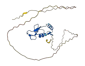
Descriptions
The autoinhibited protein was predicted that may have potential autoinhibitory elements via cis-regPred.
Autoinhibitory domains (AIDs)
Target domain |
|
Relief mechanism |
|
Assay |
cis-regPred |
Accessory elements
No accessory elements
Autoinhibited structure

Activated structure
1 structures for Q71FD5
| Entry ID | Method | Resolution | Chain | Position | Source |
|---|---|---|---|---|---|
| AF-Q71FD5-F1 | Predicted | AlphaFoldDB |
7 variants for Q71FD5
| Variant ID(s) | Position | Change | Description | Diseaes Association | Provenance |
|---|---|---|---|---|---|
| rs1132370584 | 47 | G>A | No | EVA | |
| rs258084116 | 118 | S>G | No | EVA | |
| rs3388820808 | 208 | I>K | No | EVA | |
| rs3388826917 | 213 | C>G | No | EVA | |
| rs3388831901 | 219 | K>N | No | EVA | |
| rs3388836568 | 225 | W>L | No | EVA | |
| rs3388832916 | 239 | D>Q | No | EVA |
No associated diseases with Q71FD5
4 regional properties for Q71FD5
Functions
| Description | ||
|---|---|---|
| EC Number | 2.3.2.27 | Aminoacyltransferases |
| Subcellular Localization |
|
|
| PANTHER Family | ||
| PANTHER Subfamily | ||
| PANTHER Protein Class | ||
| PANTHER Pathway Category | No pathway information available | |
13 GO annotations of cellular component
| Name | Definition |
|---|---|
| anchoring junction | A cell junction that mechanically attaches a cell (and its cytoskeleton) to neighboring cells or to the extracellular matrix. |
| cell projection | A prolongation or process extending from a cell, e.g. a flagellum or axon. |
| cytoplasm | The contents of a cell excluding the plasma membrane and nucleus, but including other subcellular structures. |
| cytoplasmic vesicle membrane | The lipid bilayer surrounding a cytoplasmic vesicle. |
| endosome membrane | The lipid bilayer surrounding an endosome. |
| fibrillar center | A structure found most metazoan nucleoli, but not usually found in lower eukaryotes; surrounded by the dense fibrillar component; the zone of transcription from multiple copies of the pre-rRNA genes is in the border region between these two structures. |
| intracellular membrane-bounded organelle | Organized structure of distinctive morphology and function, bounded by a single or double lipid bilayer membrane and occurring within the cell. Includes the nucleus, mitochondria, plastids, vacuoles, and vesicles. Excludes the plasma membrane. |
| lysosomal membrane | The lipid bilayer surrounding the lysosome and separating its contents from the cell cytoplasm. |
| membrane | A lipid bilayer along with all the proteins and protein complexes embedded in it an attached to it. |
| nucleoplasm | That part of the nuclear content other than the chromosomes or the nucleolus. |
| plasma membrane | The membrane surrounding a cell that separates the cell from its external environment. It consists of a phospholipid bilayer and associated proteins. |
| presynaptic membrane | A specialized area of membrane of the axon terminal that faces the plasma membrane of the neuron or muscle fiber with which the axon terminal establishes a synaptic junction; many synaptic junctions exhibit structural presynaptic characteristics, such as conical, electron-dense internal protrusions, that distinguish it from the remainder of the axon plasma membrane. |
| protein-containing complex | A stable assembly of two or more macromolecules, i.e. proteins, nucleic acids, carbohydrates or lipids, in which at least one component is a protein and the constituent parts function together. |
2 GO annotations of molecular function
| Name | Definition |
|---|---|
| metal ion binding | Binding to a metal ion. |
| ubiquitin protein ligase activity | Catalysis of the transfer of ubiquitin to a substrate protein via the reaction X-ubiquitin + S -> X + S-ubiquitin, where X is either an E2 or E3 enzyme, the X-ubiquitin linkage is a thioester bond, and the S-ubiquitin linkage is an amide bond: an isopeptide bond between the C-terminal glycine of ubiquitin and the epsilon-amino group of lysine residues in the substrate or, in the linear extension of ubiquitin chains, a peptide bond the between the C-terminal glycine and N-terminal methionine of ubiquitin residues. |
2 GO annotations of biological process
| Name | Definition |
|---|---|
| proteasome-mediated ubiquitin-dependent protein catabolic process | The chemical reactions and pathways resulting in the breakdown of a protein or peptide by hydrolysis of its peptide bonds, initiated by the covalent attachment of ubiquitin, and mediated by the proteasome. |
| protein K48-linked ubiquitination | A protein ubiquitination process in which a polymer of ubiquitin, formed by linkages between lysine residues at position 48 of the ubiquitin monomers, is added to a protein. K48-linked ubiquitination targets the substrate protein for degradation. |
1 homologous proteins in AiPD
| UniProt AC | Gene Name | Protein Name | Species | Evidence Code |
|---|---|---|---|---|
| Q8NHG8 | ZNRF2 | E3 ubiquitin-protein ligase ZNRF2 | Homo sapiens (Human) | PR |
| 10 | 20 | 30 | 40 | 50 | 60 |
| MGAKQSGPAA | NGRTRAYSGS | DLPSGTGSGG | GGADGARAAR | FAAPVSGAQQ | PSASAGAAAA |
| 70 | 80 | 90 | 100 | 110 | 120 |
| AAAAASAPAA | PRSRSLGGAV | GSASGGRAAQ | SAFSIPSAGG | GGGPYGSQDS | VHSSPEDSVG |
| 130 | 140 | 150 | 160 | 170 | 180 |
| ARDRDRPAGG | GPGGPRLVIG | SLPAHLSPHL | FGGFKCPVCS | KFVPSDEMDL | HLVMCLTKPR |
| 190 | 200 | 210 | 220 | 230 | |
| ITYNEDVLSK | DTGECAICLE | ELQQGDTIAR | LPCLCIYHKG | CIDEWFEVNR | SCPEHPSD |