Q6QD59
Gene name |
Bnip1 |
Protein name |
Vesicle transport protein SEC20 |
Names |
|
Species |
Mus musculus (Mouse) |
KEGG Pathway |
mmu:224630 |
EC number |
|
Protein Class |
|
Descriptions
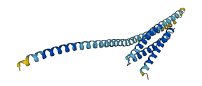
The autoinhibited protein was predicted that may have potential autoinhibitory elements via cis-regPred.
Autoinhibitory domains (AIDs)
Target domain |
|
Relief mechanism |
|
Assay |
cis-regPred |
Accessory elements
No accessory elements
Autoinhibited structure

Activated structure
1 structures for Q6QD59
| Entry ID | Method | Resolution | Chain | Position | Source |
|---|---|---|---|---|---|
| AF-Q6QD59-F1 | Predicted | AlphaFoldDB |
18 variants for Q6QD59
| Variant ID(s) | Position | Change | Description | Diseaes Association | Provenance |
|---|---|---|---|---|---|
| rs3389443956 | 4 | P>S | No | EVA | |
| rs3389443914 | 26 | L>P | No | EVA | |
| rs3389456789 | 50 | K>R | No | EVA | |
| rs3389451719 | 55 | K>E | No | EVA | |
| rs3389436282 | 77 | L>V | No | EVA | |
| rs3389436358 | 79 | Q>L | No | EVA | |
| rs3389448653 | 116 | Q>H | No | EVA | |
| rs3389451723 | 139 | T>I | No | EVA | |
| rs3389448679 | 164 | V>F | No | EVA | |
| rs3389395685 | 182 | G>E | No | EVA | |
| rs3389448707 | 186 | L>M | No | EVA | |
| rs3389436372 | 195 | N>D | No | EVA | |
| rs3389451710 | 197 | R>L | No | EVA | |
| rs3389404998 | 206 | F>I | No | EVA | |
| rs3389436353 | 209 | L>Q | No | EVA | |
| rs3389445797 | 214 | A>T | No | EVA | |
| rs3389445735 | 216 | V>F | No | EVA | |
| rs3389456757 | 227 | F>S | No | EVA |
No associated diseases with Q6QD59
No regional properties for Q6QD59
| Type | Name | Position | InterPro Accession |
|---|---|---|---|
| No domain, repeats, and functional sites for Q6QD59 | |||
Functions
9 GO annotations of cellular component
| Name | Definition |
|---|---|
| COPI-coated vesicle | A vesicle with a coat formed of the COPI coat complex proteins. COPI-coated vesicles are found associated with Golgi membranes at steady state, are involved in Golgi to endoplasmic reticulum (retrograde) vesicle transport, and possibly also in intra-Golgi transport. |
| cytoplasm | The contents of a cell excluding the plasma membrane and nucleus, but including other subcellular structures. |
| endoplasmic reticulum | The irregular network of unit membranes, visible only by electron microscopy, that occurs in the cytoplasm of many eukaryotic cells. The membranes form a complex meshwork of tubular channels, which are often expanded into slitlike cavities called cisternae. The ER takes two forms, rough (or granular), with ribosomes adhering to the outer surface, and smooth (with no ribosomes attached). |
| endoplasmic reticulum membrane | The lipid bilayer surrounding the endoplasmic reticulum. |
| integral component of membrane | The component of a membrane consisting of the gene products and protein complexes having at least some part of their peptide sequence embedded in the hydrophobic region of the membrane. |
| intracellular membrane-bounded organelle | Organized structure of distinctive morphology and function, bounded by a single or double lipid bilayer membrane and occurring within the cell. Includes the nucleus, mitochondria, plastids, vacuoles, and vesicles. Excludes the plasma membrane. |
| mitochondrial membrane | Either of the lipid bilayers that surround the mitochondrion and form the mitochondrial envelope. |
| nuclear envelope | The double lipid bilayer enclosing the nucleus and separating its contents from the rest of the cytoplasm; includes the intermembrane space, a gap of width 20-40 nm (also called the perinuclear space). |
| SNARE complex | A protein complex involved in membrane fusion; a stable ternary complex consisting of a four-helix bundle, usually formed from one R-SNARE and three Q-SNAREs with an ionic layer sandwiched between hydrophobic layers. One well-characterized example is the neuronal SNARE complex formed of synaptobrevin 2, syntaxin 1a, and SNAP-25. |
1 GO annotations of molecular function
| Name | Definition |
|---|---|
| SNAP receptor activity | Acting as a marker to identify a membrane and interacting selectively with one or more SNAREs on another membrane to mediate membrane fusion. |
5 GO annotations of biological process
| Name | Definition |
|---|---|
| apoptotic process | A programmed cell death process which begins when a cell receives an internal (e.g. DNA damage) or external signal (e.g. an extracellular death ligand), and proceeds through a series of biochemical events (signaling pathway phase) which trigger an execution phase. The execution phase is the last step of an apoptotic process, and is typically characterized by rounding-up of the cell, retraction of pseudopodes, reduction of cellular volume (pyknosis), chromatin condensation, nuclear fragmentation (karyorrhexis), plasma membrane blebbing and fragmentation of the cell into apoptotic bodies. When the execution phase is completed, the cell has died. |
| endoplasmic reticulum membrane fusion | The joining of 2 or more lipid bilayer membranes that surround the endoplasmic reticulum. |
| endoplasmic reticulum organization | A process that is carried out at the cellular level which results in the assembly, arrangement of constituent parts, or disassembly of the endoplasmic reticulum. |
| protein transport | The directed movement of proteins into, out of or within a cell, or between cells, by means of some agent such as a transporter or pore. |
| retrograde vesicle-mediated transport, Golgi to endoplasmic reticulum | The directed movement of substances from the Golgi back to the endoplasmic reticulum, mediated by vesicles bearing specific protein coats such as COPI or COG. |
1 homologous proteins in AiPD
| UniProt AC | Gene Name | Protein Name | Species | Evidence Code |
|---|---|---|---|---|
| P28791 | SEC20 | Protein transport protein SEC20 | Saccharomyces cerevisiae (strain ATCC 204508 / S288c) (Baker's yeast) | PR |
| 10 | 20 | 30 | 40 | 50 | 60 |
| MAAPQDVHVR | ICNQEIVKFD | LEVKALIQDI | RDCSGPLSEL | TELNTKVKEK | FQQLKQRIQE |
| 70 | 80 | 90 | 100 | 110 | 120 |
| LEQSAREQDK | ESEKQLLLQE | VENHKKQMLS | NQTSWRKANL | TCKLAIDNLE | KAELLQGGDS |
| 130 | 140 | 150 | 160 | 170 | 180 |
| LRQRKTTKES | LAQTSSSITE | SLMGISRMMS | QQVQQSEEAM | QTLVSSSRTL | LDANEEFKSM |
| 190 | 200 | 210 | 220 | ||
| SGTIQLGRKL | ITKYNRRELT | DKLLIFLALA | LFLATVLYIV | KKRLFPFL |