P15151
Gene name |
PVR (PVS) |
Protein name |
Poliovirus receptor |
Names |
|
Species |
Homo sapiens (Human) |
KEGG Pathway |
hsa:5817 |
EC number |
|
Protein Class |
|
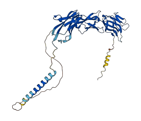
Descriptions
The autoinhibited protein was predicted that may have potential autoinhibitory elements via cis-regPred.
Autoinhibitory domains (AIDs)
Target domain |
|
Relief mechanism |
|
Assay |
cis-regPred |
Accessory elements
No accessory elements
Autoinhibited structure

Activated structure
14 structures for P15151
| Entry ID | Method | Resolution | Chain | Position | Source |
|---|---|---|---|---|---|
| 1DGI | EM | 2200 A | R | 28-329 | PDB |
| 1NN8 | EM | 1500 A | R/S/T | 28-329 | PDB |
| 3EPC | EM | 800 A | R | 30-242 | PDB |
| 3EPD | EM | 900 A | R | 30-242 | PDB |
| 3EPF | EM | 900 A | R | 30-242 | PDB |
| 3J8F | EM | 370 A | 7 | 1-417 | PDB |
| 3J9F | EM | 900 A | PDB | ||
| 3UDW | X-ray | 290 A | C/D | 28-145 | PDB |
| 3URO | X-ray | 350 A | R | 29-243 | PDB |
| 4FQP | X-ray | 360 A | A | 28-334 | PDB |
| 6ARQ | X-ray | 288 A | D | 28-334 | PDB |
| 6ISC | X-ray | 220 A | B | 28-145 | PDB |
| 6O3O | X-ray | 280 A | C/D | 28-334 | PDB |
| AF-P15151-F1 | Predicted | AlphaFoldDB |
3 variants for P15151
| Variant ID(s) | Position | Change | Description | Diseaes Association | Provenance |
|---|---|---|---|---|---|
|
rs1058402 VAR_003952 |
67 | A>T | No |
UniProt dbSNP |
|
|
VAR_049994 rs35365841 |
295 | A>T | No |
UniProt dbSNP |
|
|
rs203710 VAR_011736 |
340 | I>M | No |
UniProt dbSNP |
No associated diseases with P15151
7 regional properties for P15151
| Type | Name | Position | InterPro Accession |
|---|---|---|---|
| domain | Immunoglobulin subtype | 34 - 142 | IPR003599-1 |
| domain | Immunoglobulin subtype | 253 - 330 | IPR003599-2 |
| domain | Immunoglobulin-like domain | 24 - 123 | IPR007110-1 |
| domain | Immunoglobulin-like domain | 145 - 237 | IPR007110-2 |
| domain | Immunoglobulin-like domain | 244 - 328 | IPR007110-3 |
| domain | Immunoglobulin V-set domain | 33 - 142 | IPR013106 |
| domain | CD80-like, immunoglobulin C2-set | 148 - 231 | IPR013162 |
7 GO annotations of cellular component
| Name | Definition |
|---|---|
| adherens junction | A cell-cell junction composed of the epithelial cadherin-catenin complex. The epithelial cadherins, or E-cadherins, of each interacting cell extend through the plasma membrane into the extracellular space and bind to each other. The E-cadherins bind to catenins on the cytoplasmic side of the membrane, where the E-cadherin-catenin complex binds to cytoskeletal components and regulatory and signaling molecules. |
| cell surface | The external part of the cell wall and/or plasma membrane. |
| cytoplasm | The contents of a cell excluding the plasma membrane and nucleus, but including other subcellular structures. |
| extracellular space | That part of a multicellular organism outside the cells proper, usually taken to be outside the plasma membranes, and occupied by fluid. |
| focal adhesion | A cell-substrate junction that anchors the cell to the extracellular matrix and that forms a point of termination of actin filaments. In insects focal adhesion has also been referred to as hemi-adherens junction (HAJ). |
| integral component of membrane | The component of a membrane consisting of the gene products and protein complexes having at least some part of their peptide sequence embedded in the hydrophobic region of the membrane. |
| plasma membrane | The membrane surrounding a cell that separates the cell from its external environment. It consists of a phospholipid bilayer and associated proteins. |
3 GO annotations of molecular function
| Name | Definition |
|---|---|
| cell adhesion molecule binding | Binding to a cell adhesion molecule. |
| signaling receptor activity | Receiving a signal and transmitting it in the cell to initiate a change in cell activity. A signal is a physical entity or change in state that is used to transfer information in order to trigger a response. |
| virus receptor activity | Combining with a virus component and mediating entry of the virus into the cell. |
6 GO annotations of biological process
| Name | Definition |
|---|---|
| heterophilic cell-cell adhesion via plasma membrane cell adhesion molecules | The attachment of an adhesion molecule in one cell to a nonidentical adhesion molecule in an adjacent cell. |
| homophilic cell adhesion via plasma membrane adhesion molecules | The attachment of a plasma membrane adhesion molecule in one cell to an identical molecule in an adjacent cell. |
| positive regulation of natural killer cell mediated cytotoxicity | Any process that activates or increases the frequency, rate or extent of natural killer cell mediated cytotoxicity. |
| positive regulation of natural killer cell mediated cytotoxicity directed against tumor cell target | Any process that activates or increases the frequency, rate, or extent of natural killer cell mediated cytotoxicity directed against tumor cell target. |
| susceptibility to natural killer cell mediated cytotoxicity | The process of causing a cell to become susceptible to natural killer cell mediated cytotoxicity. |
| susceptibility to T cell mediated cytotoxicity | The process of causing a cell to become susceptible to T cell mediated cytotoxicity. |
1 homologous proteins in AiPD
| UniProt AC | Gene Name | Protein Name | Species | Evidence Code |
|---|---|---|---|---|
| P32507 | Nectin2 | Nectin-2 | Mus musculus (Mouse) | PR |
| 10 | 20 | 30 | 40 | 50 | 60 |
| MARAMAAAWP | LLLVALLVLS | WPPPGTGDVV | VQAPTQVPGF | LGDSVTLPCY | LQVPNMEVTH |
| 70 | 80 | 90 | 100 | 110 | 120 |
| VSQLTWARHG | ESGSMAVFHQ | TQGPSYSESK | RLEFVAARLG | AELRNASLRM | FGLRVEDEGN |
| 130 | 140 | 150 | 160 | 170 | 180 |
| YTCLFVTFPQ | GSRSVDIWLR | VLAKPQNTAE | VQKVQLTGEP | VPMARCVSTG | GRPPAQITWH |
| 190 | 200 | 210 | 220 | 230 | 240 |
| SDLGGMPNTS | QVPGFLSGTV | TVTSLWILVP | SSQVDGKNVT | CKVEHESFEK | PQLLTVNLTV |
| 250 | 260 | 270 | 280 | 290 | 300 |
| YYPPEVSISG | YDNNWYLGQN | EATLTCDARS | NPEPTGYNWS | TTMGPLPPFA | VAQGAQLLIR |
| 310 | 320 | 330 | 340 | 350 | 360 |
| PVDKPINTTL | ICNVTNALGA | RQAELTVQVK | EGPPSEHSGI | SRNAIIFLVL | GILVFLILLG |
| 370 | 380 | 390 | 400 | 410 | |
| IGIYFYWSKC | SREVLWHCHL | CPSSTEHASA | SANGHVSYSA | VSRENSSSQD | PQTEGTR |