Q9P4X1
Gene name |
SPAC959.05c |
Protein name |
Thioredoxin domain-containing protein C959.05c |
Names |
Putative protein disulfide isomerase C959.05c |
Species |
Schizosaccharomyces pombe (strain 972 / ATCC 24843) (Fission yeast) |
KEGG Pathway |
spo:SPAC959.05c |
EC number |
5.3.4.1: Transposing S-S bonds |
Protein Class |
|
Descriptions
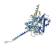
The autoinhibited protein was predicted that may have potential autoinhibitory elements via cis-regPred.
Autoinhibitory domains (AIDs)
Target domain |
|
Relief mechanism |
|
Assay |
cis-regPred |
Accessory elements
No accessory elements
Autoinhibited structure

Activated structure
1 structures for Q9P4X1
| Entry ID | Method | Resolution | Chain | Position | Source |
|---|---|---|---|---|---|
| AF-Q9P4X1-F1 | Predicted | AlphaFoldDB |
16 variants for Q9P4X1
| Variant ID(s) | Position | Change | Description | Diseaes Association | Provenance |
|---|---|---|---|---|---|
| I_3397586_C_T | 18 | S>N | No | Jeffares_SNPs | |
| I_3397472_T_C | 56 | D>G | No | Jeffares_SNPs | |
| I_3397246_T_C | 113 | E>G | No | Jeffares_SNPs | |
| I_3397157_C_T | 143 | D>N | No | Jeffares_SNPs | |
| I_3397117_C_T | 156 | S>N | No | Jeffares_SNPs | |
| I_3397093_A_G | 164 | L>P | No | Jeffares_SNPs | |
| I_3396608_T_C | 326 | K>E | No | Jeffares_SNPs | |
| I_3396523_C_T | 354 | S>N | No | Jeffares_SNPs | |
| I_3396403_T_G | 394 | E>A | No | Jeffares_SNPs | |
| I_3396264_C_T | 440 | M>I | No | Jeffares_SNPs | |
| I_3396080_C_T | 502 | V>M | No | Jeffares_SNPs | |
| I_3395911_G_A | 558 | S>L | No | Jeffares_SNPs | |
| I_3395891_G_A | 565 | R>C | No | Jeffares_SNPs | |
| I_3395881_C_T | 568 | G>D | No | Jeffares_SNPs | |
| I_3395825_T_A | 587 | N>Y | No | Jeffares_SNPs | |
| I_3395714_G_C | 624 | H>D | No | Jeffares_SNPs |
No associated diseases with Q9P4X1
1 regional properties for Q9P4X1
| Type | Name | Position | InterPro Accession |
|---|---|---|---|
| domain | Thioredoxin domain | 153 - 284 | IPR013766 |
Functions
| Description | ||
|---|---|---|
| EC Number | 5.3.4.1 | Transposing S-S bonds |
| Subcellular Localization |
|
|
| PANTHER Family | ||
| PANTHER Subfamily | ||
| PANTHER Protein Class | ||
| PANTHER Pathway Category | No pathway information available | |
4 GO annotations of cellular component
| Name | Definition |
|---|---|
| endoplasmic reticulum | The irregular network of unit membranes, visible only by electron microscopy, that occurs in the cytoplasm of many eukaryotic cells. The membranes form a complex meshwork of tubular channels, which are often expanded into slitlike cavities called cisternae. The ER takes two forms, rough (or granular), with ribosomes adhering to the outer surface, and smooth (with no ribosomes attached). |
| endoplasmic reticulum lumen | The volume enclosed by the membranes of the endoplasmic reticulum. |
| endoplasmic reticulum membrane | The lipid bilayer surrounding the endoplasmic reticulum. |
| integral component of membrane | The component of a membrane consisting of the gene products and protein complexes having at least some part of their peptide sequence embedded in the hydrophobic region of the membrane. |
2 GO annotations of molecular function
| Name | Definition |
|---|---|
| protein disulfide isomerase activity | Catalysis of the rearrangement of both intrachain and interchain disulfide bonds in proteins. |
| protein-disulfide reductase activity | Catalysis of the reaction: a protein with reduced sulfide groups = a protein with oxidized disulfide bonds. |
4 GO annotations of biological process
| Name | Definition |
|---|---|
| polyamine metabolic process | The chemical reactions and pathways involving polyamines, any organic compound containing two or more amino groups. |
| protein retention in ER lumen | The retention in the endoplasmic reticulum (ER) lumen of soluble resident proteins. Sorting receptors retrieve proteins with ER localization signals, such as KDEL and HDEL sequences or some transmembrane domains, that have escaped to the cis-Golgi network and return them to the ER. Abnormally folded proteins and unassembled subunits are also selectively retained in the ER. |
| response to endoplasmic reticulum stress | Any process that results in a change in state or activity of a cell (in terms of movement, secretion, enzyme production, gene expression, etc.) as a result of a stress acting at the endoplasmic reticulum. ER stress usually results from the accumulation of unfolded or misfolded proteins in the ER lumen. |
| ubiquitin-dependent ERAD pathway | The series of steps necessary to target endoplasmic reticulum (ER)-resident proteins for degradation by the cytoplasmic proteasome. Begins with recognition of the ER-resident protein, includes retrotranslocation (dislocation) of the protein from the ER to the cytosol, protein ubiquitination necessary for correct substrate transfer, transport of the protein to the proteasome, and ends with degradation of the protein by the cytoplasmic proteasome. |
No homologous proteins in AiPD
| UniProt AC | Gene Name | Protein Name | Species | Evidence Code |
|---|---|---|---|---|
| No homologous proteins | ||||
| 10 | 20 | 30 | 40 | 50 | 60 |
| MKLFLYHFTF | IVYYFIISFS | YAFSIKQEII | VSSHNASSIL | NTTAFWFVEF | TESKYDKEEF |
| 70 | 80 | 90 | 100 | 110 | 120 |
| SVIWNEVSME | FPDIRRAKVF | CDLDLEFCAQ | QEIYDHPKVV | VLKNGMWMRH | VLEKNQITTK |
| 130 | 140 | 150 | 160 | 170 | 180 |
| SARQFVKSHL | GNCDLEQAEN | ETDCFSDDGE | YNSDSSSTDP | AFELKEDQSW | KHSSILRPLE |
| 190 | 200 | 210 | 220 | 230 | 240 |
| TLNFKRFLFG | NEIMSKTRAF | VMFVSLKHCE | DCFHWEAVWS | SITRNTDERL | KMAQVNCDEE |
| 250 | 260 | 270 | 280 | 290 | 300 |
| KEMCNHFHIK | KFPTFRVFQG | FDSIQYNGPL | KYQQLLSYSN | QVASYQAIKI | EEGDIESIEN |
| 310 | 320 | 330 | 340 | 350 | 360 |
| SHPVFFLVLY | DFATTSEDFS | IIERLKLQLA | GVAPLYICNS | KALANKYGAQ | SQPSIIAVRN |
| 370 | 380 | 390 | 400 | 410 | 420 |
| GMPIVYQAIT | PREFRDYKRI | TEWINIVSSP | FITELTPTKC | HSLLNRKLTV | LTLLQPDSEQ |
| 430 | 440 | 450 | 460 | 470 | 480 |
| FFSSQEELLR | LGKRWFRFQM | QRQRNDIVWS | RIKKYSAIAE | AKKKGFARKV | KRIKYSKISH |
| 490 | 500 | 510 | 520 | 530 | 540 |
| PTYTESVSFL | WLDSSLWLDW | IVENLDHTVY | VDSITPPVFV | IDHSKGVIYV | SDRNGNSLTL |
| 550 | 560 | 570 | 580 | 590 | 600 |
| EEDSLFSTLR | IILEHPNSSR | LQKLRAPGLC | PNGSPNYRNR | YKLIVFNLLI | ALLILSILTI |
| 610 | 620 | 630 | |||
| ISASRLSRRR | RQLLNKQPVF | GFYHSLVIAK | SD |