Q99L04
Gene name |
Dhrs1 |
Protein name |
Dehydrogenase/reductase SDR family member 1 |
Names |
Short chain dehydrogenase/reductase family 19C member 1, Protein SDR19C1 |
Species |
Mus musculus (Mouse) |
KEGG Pathway |
mmu:52585 |
EC number |
|
Protein Class |
|
Descriptions
The autoinhibited protein was predicted that may have potential autoinhibitory elements via cis-regPred.
Autoinhibitory domains (AIDs)
Target domain |
|
Relief mechanism |
|
Assay |
cis-regPred |
Accessory elements
No accessory elements
Autoinhibited structure

Activated structure
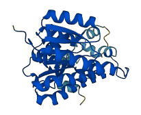
1 structures for Q99L04
| Entry ID | Method | Resolution | Chain | Position | Source |
|---|---|---|---|---|---|
| AF-Q99L04-F1 | Predicted | AlphaFoldDB |
11 variants for Q99L04
| Variant ID(s) | Position | Change | Description | Diseaes Association | Provenance |
|---|---|---|---|---|---|
| rs3389304780 | 38 | G>S | No | EVA | |
| rs3389321252 | 72 | K>R | No | EVA | |
| rs3389254311 | 82 | Q>K | No | EVA | |
| rs3389327248 | 137 | L>V | No | EVA | |
| rs3389341611 | 143 | K>E | No | EVA | |
| rs3389330551 | 178 | A>P | No | EVA | |
| rs3389333453 | 179 | H>R | No | EVA | |
| rs3389314568 | 199 | E>G | No | EVA | |
| rs3389334751 | 261 | R>C | No | EVA | |
| rs3389327226 | 271 | P>H | No | EVA | |
| rs3389333429 | 297 | G>D | No | EVA |
No associated diseases with Q99L04
7 regional properties for Q99L04
| Type | Name | Position | InterPro Accession |
|---|---|---|---|
| domain | BRCT domain | 27 - 124 | IPR001357 |
| domain | DNA-directed DNA polymerase X | 163 - 508 | IPR002054 |
| domain | DNA polymerase beta-like, N-terminal domain | 166 - 230 | IPR010996 |
| domain | DNA polymerase lambda, fingers domain | 250 - 299 | IPR018944 |
| binding_site | DNA polymerase family X, binding site | 332 - 351 | IPR019843 |
| domain | DNA polymerase beta, palm domain | 305 - 375 | IPR028207 |
| domain | DNA polymerase beta, thumb domain | 445 - 508 | IPR029398 |
3 GO annotations of cellular component
| Name | Definition |
|---|---|
| endoplasmic reticulum | The irregular network of unit membranes, visible only by electron microscopy, that occurs in the cytoplasm of many eukaryotic cells. The membranes form a complex meshwork of tubular channels, which are often expanded into slitlike cavities called cisternae. The ER takes two forms, rough (or granular), with ribosomes adhering to the outer surface, and smooth (with no ribosomes attached). |
| mitochondrial inner membrane | The inner, i.e. lumen-facing, lipid bilayer of the mitochondrial envelope. It is highly folded to form cristae. |
| mitochondrion | A semiautonomous, self replicating organelle that occurs in varying numbers, shapes, and sizes in the cytoplasm of virtually all eukaryotic cells. It is notably the site of tissue respiration. |
3 GO annotations of molecular function
| Name | Definition |
|---|---|
| carbonyl reductase (NADPH) activity | Catalysis of the reaction: R-CHOH-R' + NADP+ = R-CO-R' + NADPH + H+. |
| oxidoreductase activity, acting on the CH-OH group of donors, NAD or NADP as acceptor | Catalysis of an oxidation-reduction (redox) reaction in which a CH-OH group acts as a hydrogen or electron donor and reduces NAD+ or NADP. |
| testosterone 17-beta-dehydrogenase (NADP+) activity | Catalysis of the reaction: NADP+ + testosterone = NADPH + H+ + androst-4-ene-3,17-dione. |
No GO annotations of biological process
| Name | Definition |
|---|---|
| No GO annotations for biological process |
3 homologous proteins in AiPD
| UniProt AC | Gene Name | Protein Name | Species | Evidence Code |
|---|---|---|---|---|
| Q9BTZ2 | DHRS4 | Dehydrogenase/reductase SDR family member 4 | Homo sapiens (Human) | PR |
| Q96LJ7 | DHRS1 | Dehydrogenase/reductase SDR family member 1 | Homo sapiens (Human) | PR |
| Q84ST4 | NOL | Chlorophyll(ide) b reductase NOL, chloroplastic | Oryza sativa subsp japonica (Rice) | PR |
| 10 | 20 | 30 | 40 | 50 | 60 |
| MVAPMKGQVC | VVTGASRGIG | RGIALQLCKA | GATVYITGRH | LDTLRATAQE | AQSLGGRCVP |
| 70 | 80 | 90 | 100 | 110 | 120 |
| VVCDSSQESE | VKSLFEQVDR | EQKGRLDVLV | NNAYAGVQAI | LNTTNKSFWE | VPASIWDDIN |
| 130 | 140 | 150 | 160 | 170 | 180 |
| NVGLRGHYLC | SVYGARLMVP | AGKGLIVIVS | SPGGLQHMFN | VPYGVGKAAC | DRLAADCAHE |
| 190 | 200 | 210 | 220 | 230 | 240 |
| LRRHGVSYVS | LWPGLVQTEM | VKEFMAKEDT | PEDPLFKKMK | PDFSSAESPE | MSGKCVVALA |
| 250 | 260 | 270 | 280 | 290 | 300 |
| TDPNILNLSG | KVLPSCDLAR | RYGLKDIDGR | PVKDYFSLGY | ALSQVSSLGW | LNSFLPGFLR |
| 310 | |||||
| VPKWVVTLYN | SKF |