Q921V5
Gene name |
Mgat2 |
Protein name |
Alpha-1,6-mannosyl-glycoprotein 2-beta-N-acetylglucosaminyltransferase |
Names |
Beta-1,2-N-acetylglucosaminyltransferase II, GlcNAc-T II, GNT-II, Mannoside acetylglucosaminyltransferase 2, N-glycosyl-oligosaccharide-glycoprotein N-acetylglucosaminyltransferase II |
Species |
Mus musculus (Mouse) |
KEGG Pathway |
mmu:217664 |
EC number |
2.4.1.143: Hexosyltransferases |
Protein Class |
|
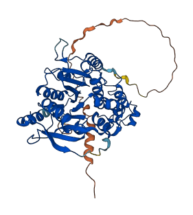
Descriptions
The autoinhibited protein was predicted that may have potential autoinhibitory elements via cis-regPred.
Autoinhibitory domains (AIDs)
Target domain |
|
Relief mechanism |
|
Assay |
cis-regPred |
Accessory elements
No accessory elements
Autoinhibited structure

Activated structure
1 structures for Q921V5
| Entry ID | Method | Resolution | Chain | Position | Source |
|---|---|---|---|---|---|
| AF-Q921V5-F1 | Predicted | AlphaFoldDB |
7 variants for Q921V5
| Variant ID(s) | Position | Change | Description | Diseaes Association | Provenance |
|---|---|---|---|---|---|
| rs586106993 | 73 | A>G | No | EVA | |
| rs3403006849 | 120 | H>P | No | EVA | |
| rs3403394495 | 175 | S>R | No | EVA | |
| rs3403575605 | 253 | L>Q | No | EVA | |
| rs3403315906 | 256 | D>G | No | EVA | |
| rs3389249826 | 287 | G>E | No | EVA | |
| rs3389215952 | 294 | S>I | No | EVA |
No associated diseases with Q921V5
No regional properties for Q921V5
| Type | Name | Position | InterPro Accession |
|---|---|---|---|
| No domain, repeats, and functional sites for Q921V5 | |||
Functions
| Description | ||
|---|---|---|
| EC Number | 2.4.1.143 | Hexosyltransferases |
| Subcellular Localization |
|
|
| PANTHER Family | ||
| PANTHER Subfamily | ||
| PANTHER Protein Class | ||
| PANTHER Pathway Category | No pathway information available | |
4 GO annotations of cellular component
| Name | Definition |
|---|---|
| Golgi apparatus | A membrane-bound cytoplasmic organelle of the endomembrane system that further processes the core oligosaccharides (e.g. N-glycans) added to proteins in the endoplasmic reticulum and packages them into membrane-bound vesicles. The Golgi apparatus operates at the intersection of the secretory, lysosomal, and endocytic pathways. |
| Golgi membrane | The lipid bilayer surrounding any of the compartments of the Golgi apparatus. |
| Golgi stack | The set of thin, flattened membrane-bounded compartments, called cisternae, that form the central portion of the Golgi complex. The stack usually comprises cis, medial, and trans cisternae; the cis- and trans-Golgi networks are not considered part of the stack. |
| integral component of membrane | The component of a membrane consisting of the gene products and protein complexes having at least some part of their peptide sequence embedded in the hydrophobic region of the membrane. |
4 GO annotations of molecular function
| Name | Definition |
|---|---|
| alpha-1,6-mannosylglycoprotein 2-beta-N-acetylglucosaminyltransferase activity | Catalysis of the reaction: UDP-N-acetyl-D-glucosamine + alpha-D-mannosyl-1,6-(N-acetyl-beta-D-glucosaminyl-1,2-alpha-D-mannosyl-1,3)-beta-D-mannosyl-R = UDP + N-acetyl-beta-D-glucosaminyl-1,2-alpha-D-mannosyl-1,6-(N-acetyl-beta-D-glucosaminyl-1,2-alpha-D-mannosyl-1,3)-beta-D-mannosyl-R. |
| carbohydrate binding | Binding to a carbohydrate, which includes monosaccharides, oligosaccharides and polysaccharides as well as substances derived from monosaccharides by reduction of the carbonyl group (alditols), by oxidation of one or more hydroxy groups to afford the corresponding aldehydes, ketones, or carboxylic acids, or by replacement of one or more hydroxy group(s) by a hydrogen atom. Cyclitols are generally not regarded as carbohydrates. |
| manganese ion binding | Binding to a manganese ion (Mn). |
| protein homodimerization activity | Binding to an identical protein to form a homodimer. |
3 GO annotations of biological process
| Name | Definition |
|---|---|
| oligosaccharide biosynthetic process | The chemical reactions and pathways resulting in the formation of oligosaccharides, molecules with between two and (about) 20 monosaccharide residues connected by glycosidic linkages. |
| protein N-linked glycosylation | A protein glycosylation process in which a carbohydrate or carbohydrate derivative unit is added to a protein via the N4 atom of peptidyl-asparagine, the omega-N of arginine, or the N1' atom peptidyl-tryptophan. |
| protein N-linked glycosylation via asparagine | The glycosylation of protein via the N4 atom of peptidyl-asparagine forming N4-glycosyl-L-asparagine; the most common form is N-acetylglucosaminyl asparagine; N-acetylgalactosaminyl asparagine and N4 glucosyl asparagine also occur. This modification typically occurs in extracellular peptides with an N-X-(ST) motif. Partial modification has been observed to occur with cysteine, rather than serine or threonine, in the third position; secondary structure features are important, and proline in the second or fourth positions inhibits modification. |
3 homologous proteins in AiPD
| UniProt AC | Gene Name | Protein Name | Species | Evidence Code |
|---|---|---|---|---|
| Q10469 | MGAT2 | Alpha-1,6-mannosyl-glycoprotein 2-beta-N-acetylglucosaminyltransferase | Homo sapiens (Human) | PR |
| O19071 | MGAT2 | Alpha-1,6-mannosyl-glycoprotein 2-beta-N-acetylglucosaminyltransferase | Sus scrofa (Pig) | PR |
| Q09326 | Mgat2 | Alpha-1,6-mannosyl-glycoprotein 2-beta-N-acetylglucosaminyltransferase | Rattus norvegicus (Rat) | PR |
| 10 | 20 | 30 | 40 | 50 | 60 |
| MRFRIYKRKV | LILTLVVAAC | GFVLWSSNGR | QRKSDALGPP | LLDAEPVRGA | GHLAVSVGIR |
| 70 | 80 | 90 | 100 | 110 | 120 |
| RVSNESAAPL | VPAVPRPEVD | NLTLRYRSLV | YQLNFDQMLR | NVGNDGTWSP | GELVLVVQVH |
| 130 | 140 | 150 | 160 | 170 | 180 |
| NRPEYLRLLI | DSLRKAQGIQ | EVLVIFSHDF | WSAEINSLIS | RVDFCPVLQV | FFPFSIQLYP |
| 190 | 200 | 210 | 220 | 230 | 240 |
| NEFPGSDPRD | CPRDLKKNAA | LKLGCINAEY | PDSFGHYREA | KFSQTKHHWW | WKLHFVWERV |
| 250 | 260 | 270 | 280 | 290 | 300 |
| KVLQDYTGLI | LFLEEDHYLA | PDFYHVFKKM | WKLKQQECPG | CDVLSLGTYT | TIRSFYGIAD |
| 310 | 320 | 330 | 340 | 350 | 360 |
| KVDVKTWKST | EHNMGLALTR | DAYQKLIECT | DTFCTYDDYN | WDWTLQYLTL | ACLPKIWKVL |
| 370 | 380 | 390 | 400 | 410 | 420 |
| VPQAPRIFHA | GDCGMHHKKT | CRPSTQSAQI | ESLLNSNKQY | LFPETLVIGE | KFPMAAISPP |
| 430 | 440 | ||||
| RKNGGWGDIR | DHELCKSYRR | LQ |