Q5RB77
Gene name |
PDCL3 |
Protein name |
Phosducin-like protein 3 |
Names |
|
Species |
Pongo abelii (Sumatran orangutan) (Pongo pygmaeus abelii) |
KEGG Pathway |
pon:100172481 |
EC number |
|
Protein Class |
|
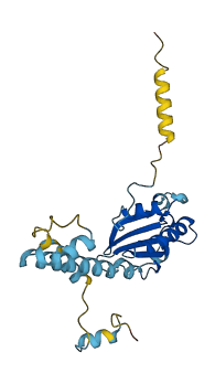
Descriptions
The autoinhibited protein was predicted that may have potential autoinhibitory elements via cis-regPred.
Autoinhibitory domains (AIDs)
Target domain |
|
Relief mechanism |
|
Assay |
cis-regPred |
Accessory elements
No accessory elements
Autoinhibited structure

Activated structure
1 structures for Q5RB77
| Entry ID | Method | Resolution | Chain | Position | Source |
|---|---|---|---|---|---|
| AF-Q5RB77-F1 | Predicted | AlphaFoldDB |
No variants for Q5RB77
| Variant ID(s) | Position | Change | Description | Diseaes Association | Provenance |
|---|---|---|---|---|---|
| No variants for Q5RB77 | |||||
No associated diseases with Q5RB77
1 regional properties for Q5RB77
| Type | Name | Position | InterPro Accession |
|---|---|---|---|
| domain | Phosducin, thioredoxin-like domain | 32 - 180 | IPR024253 |
2 GO annotations of cellular component
| Name | Definition |
|---|---|
| endoplasmic reticulum | The irregular network of unit membranes, visible only by electron microscopy, that occurs in the cytoplasm of many eukaryotic cells. The membranes form a complex meshwork of tubular channels, which are often expanded into slitlike cavities called cisternae. The ER takes two forms, rough (or granular), with ribosomes adhering to the outer surface, and smooth (with no ribosomes attached). |
| perinuclear region of cytoplasm | Cytoplasm situated near, or occurring around, the nucleus. |
No GO annotations of molecular function
| Name | Definition |
|---|---|
| No GO annotations for molecular function |
2 GO annotations of biological process
| Name | Definition |
|---|---|
| angiogenesis | Blood vessel formation when new vessels emerge from the proliferation of pre-existing blood vessels. |
| apoptotic process | A programmed cell death process which begins when a cell receives an internal (e.g. DNA damage) or external signal (e.g. an extracellular death ligand), and proceeds through a series of biochemical events (signaling pathway phase) which trigger an execution phase. The execution phase is the last step of an apoptotic process, and is typically characterized by rounding-up of the cell, retraction of pseudopodes, reduction of cellular volume (pyknosis), chromatin condensation, nuclear fragmentation (karyorrhexis), plasma membrane blebbing and fragmentation of the cell into apoptotic bodies. When the execution phase is completed, the cell has died. |
No homologous proteins in AiPD
| UniProt AC | Gene Name | Protein Name | Species | Evidence Code |
|---|---|---|---|---|
| No homologous proteins | ||||
| 10 | 20 | 30 | 40 | 50 | 60 |
| MQDPNADTEW | NDILRKKGIL | PPKESLKELE | EEAEEEQRIL | QQSVVKTYED | MTLEELDDHE |
| 70 | 80 | 90 | 100 | 110 | 120 |
| DEFNEEDERA | IEMYRRQRLA | EWKVTKLKNK | FGEVLEISGK | DYVQEVTKAG | EGLWVILHLY |
| 130 | 140 | 150 | 160 | 170 | 180 |
| KQGIPLCALI | NQHLSGLARK | FPDVKFIKAI | STTCIPNYPD | RNLPTIFVYL | EGDIKAQFIG |
| 190 | 200 | 210 | 220 | 230 | |
| PLVFGGMNLT | RDELEWKLSE | SGAIMTDLEE | NPKKPIEDVL | LSSVRRSALM | KRDSDSEGD |