Q23858
Gene name |
crtA (DDB_G0283539) |
Protein name |
Calreticulin |
Names |
|
Species |
Dictyostelium discoideum (Slime mold) |
KEGG Pathway |
ddi:DDB_G0283539 |
EC number |
|
Protein Class |
|
Descriptions
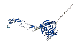
The autoinhibited protein was predicted that may have potential autoinhibitory elements via cis-regPred.
Autoinhibitory domains (AIDs)
Target domain |
|
Relief mechanism |
|
Assay |
cis-regPred |
Accessory elements
No accessory elements
Autoinhibited structure

Activated structure
1 structures for Q23858
| Entry ID | Method | Resolution | Chain | Position | Source |
|---|---|---|---|---|---|
| AF-Q23858-F1 | Predicted | AlphaFoldDB |
No variants for Q23858
| Variant ID(s) | Position | Change | Description | Diseaes Association | Provenance |
|---|---|---|---|---|---|
| No variants for Q23858 | |||||
No associated diseases with Q23858
14 regional properties for Q23858
| Type | Name | Position | InterPro Accession |
|---|---|---|---|
| repeat | HAT (Half-A-TPR) repeat | 55 - 87 | IPR003107-1 |
| repeat | HAT (Half-A-TPR) repeat | 89 - 121 | IPR003107-2 |
| repeat | HAT (Half-A-TPR) repeat | 123 - 155 | IPR003107-3 |
| repeat | HAT (Half-A-TPR) repeat | 157 - 188 | IPR003107-4 |
| repeat | HAT (Half-A-TPR) repeat | 190 - 221 | IPR003107-5 |
| repeat | HAT (Half-A-TPR) repeat | 223 - 258 | IPR003107-6 |
| repeat | HAT (Half-A-TPR) repeat | 260 - 294 | IPR003107-7 |
| repeat | HAT (Half-A-TPR) repeat | 304 - 336 | IPR003107-8 |
| repeat | HAT (Half-A-TPR) repeat | 338 - 372 | IPR003107-9 |
| repeat | HAT (Half-A-TPR) repeat | 382 - 418 | IPR003107-10 |
| repeat | HAT (Half-A-TPR) repeat | 420 - 451 | IPR003107-11 |
| repeat | HAT (Half-A-TPR) repeat | 453 - 485 | IPR003107-12 |
| repeat | HAT (Half-A-TPR) repeat | 487 - 521 | IPR003107-13 |
| repeat | HAT (Half-A-TPR) repeat | 523 - 554 | IPR003107-14 |
4 GO annotations of cellular component
| Name | Definition |
|---|---|
| endoplasmic reticulum | The irregular network of unit membranes, visible only by electron microscopy, that occurs in the cytoplasm of many eukaryotic cells. The membranes form a complex meshwork of tubular channels, which are often expanded into slitlike cavities called cisternae. The ER takes two forms, rough (or granular), with ribosomes adhering to the outer surface, and smooth (with no ribosomes attached). |
| endoplasmic reticulum lumen | The volume enclosed by the membranes of the endoplasmic reticulum. |
| endoplasmic reticulum membrane | The lipid bilayer surrounding the endoplasmic reticulum. |
| phagocytic vesicle | A membrane-bounded intracellular vesicle that arises from the ingestion of particulate material by phagocytosis. |
3 GO annotations of molecular function
| Name | Definition |
|---|---|
| calcium ion binding | Binding to a calcium ion (Ca2+). |
| carbohydrate binding | Binding to a carbohydrate, which includes monosaccharides, oligosaccharides and polysaccharides as well as substances derived from monosaccharides by reduction of the carbonyl group (alditols), by oxidation of one or more hydroxy groups to afford the corresponding aldehydes, ketones, or carboxylic acids, or by replacement of one or more hydroxy group(s) by a hydrogen atom. Cyclitols are generally not regarded as carbohydrates. |
| unfolded protein binding | Binding to an unfolded protein. |
4 GO annotations of biological process
| Name | Definition |
|---|---|
| phagocytosis, engulfment | The internalization of bacteria, immune complexes and other particulate matter or of an apoptotic cell by phagocytosis, including the membrane and cytoskeletal processes required, which involves one of three mechanisms: zippering of pseudopods around a target via repeated receptor-ligand interactions, sinking of the target directly into plasma membrane of the phagocytosing cell, or induced uptake via an enhanced membrane ruffling of the phagocytosing cell similar to macropinocytosis. |
| protein folding | The process of assisting in the covalent and noncovalent assembly of single chain polypeptides or multisubunit complexes into the correct tertiary structure. |
| response to other organism | Any process that results in a change in state or activity of a cell or an organism (in terms of movement, secretion, enzyme production, gene expression, etc.) as a result of a stimulus from another living organism. |
| ubiquitin-dependent ERAD pathway | The series of steps necessary to target endoplasmic reticulum (ER)-resident proteins for degradation by the cytoplasmic proteasome. Begins with recognition of the ER-resident protein, includes retrotranslocation (dislocation) of the protein from the ER to the cytosol, protein ubiquitination necessary for correct substrate transfer, transport of the protein to the proteasome, and ends with degradation of the protein by the cytoplasmic proteasome. |
No homologous proteins in AiPD
| UniProt AC | Gene Name | Protein Name | Species | Evidence Code |
|---|---|---|---|---|
| No homologous proteins | ||||
| 10 | 20 | 30 | 40 | 50 | 60 |
| MRLLLCLIFL | VFVFNFALST | VHFKDTFDND | WESRWVVSDW | HKEDGKSGKL | VHTAGKWFGD |
| 70 | 80 | 90 | 100 | 110 | 120 |
| ENQKGIQTSE | DARFYAVSAK | FPSFSNKGKD | LVLQYTVKNE | QKVDCGGSYI | KLLPSKLDQS |
| 130 | 140 | 150 | 160 | 170 | 180 |
| AFDGESEYSI | MFGPDVCGAS | KRVHVILNYK | GKNHLIKKEI | NKVETDQLTH | QYTLVISPDN |
| 190 | 200 | 210 | 220 | 230 | 240 |
| TYKVLVDNKE | IQAGNLADDW | ELLPSKQIKD | PKQSKPVDWV | DVKEIDDPED | VKPAGHDDIP |
| 250 | 260 | 270 | 280 | 290 | 300 |
| ASIVDPEAVK | PEDWNEEDDG | EWEAPTIANP | EYKGEWKAKK | IPNPEYKGEW | VHPLIDNPEY |
| 310 | 320 | 330 | 340 | 350 | 360 |
| AEDNELYLFN | DLGAIGFELW | QVKSGSIFNN | MIVTDSVEEA | KDFSEKTFVA | NQEAEKKMFD |
| 370 | 380 | 390 | 400 | 410 | 420 |
| DLEAAKAEER | KKADEKLAAE | KAAEKEAEEA | DEEEEEVAEE | DLVKTDDKKE | EVKKSTKKVD |
| HDEL |