Q10159
Gene name |
myh1 (myh, SPAC26A3.02) |
Protein name |
Adenine DNA glycosylase |
Names |
|
Species |
Schizosaccharomyces pombe (strain 972 / ATCC 24843) (Fission yeast) |
KEGG Pathway |
spo:SPAC26A3.02 |
EC number |
3.2.2.31: Hydrolyzing N-glycosyl compounds |
Protein Class |
|
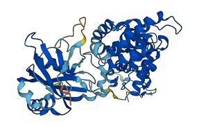
Descriptions
The autoinhibited protein was predicted that may have potential autoinhibitory elements via cis-regPred.
Autoinhibitory domains (AIDs)
Target domain |
|
Relief mechanism |
|
Assay |
cis-regPred |
Accessory elements
No accessory elements
Autoinhibited structure

Activated structure
1 structures for Q10159
| Entry ID | Method | Resolution | Chain | Position | Source |
|---|---|---|---|---|---|
| AF-Q10159-F1 | Predicted | AlphaFoldDB |
6 variants for Q10159
| Variant ID(s) | Position | Change | Description | Diseaes Association | Provenance |
|---|---|---|---|---|---|
| I_3335040_G_A | 17 | E>K | No | Jeffares_SNPs | |
| I_3335170_A_G | 60 | Q>R | No | Jeffares_SNPs | |
| I_3336002_A_G | 290 | H>R | No | Jeffares_SNPs | |
| I_3336316_A_G | 395 | I>V | No | Jeffares_SNPs | |
| I_3336487_G_A | 452 | A>T | No | Jeffares_SNPs | |
| I_3336499_G_A | 456 | V>I | No | Jeffares_SNPs |
No associated diseases with Q10159
6 regional properties for Q10159
| Type | Name | Position | InterPro Accession |
|---|---|---|---|
| conserved_site | Helix-hairpin-helix motif | 134 - 162 | IPR000445 |
| domain | HhH-GPD domain | 63 - 224 | IPR003265 |
| conserved_site | Endonuclease III-like, iron-sulphur cluster loop motif | 225 - 245 | IPR003651 |
| binding_site | Endonuclease III, iron-sulphur binding site | 226 - 242 | IPR004035 |
| conserved_site | Endonuclease III-like, conserved site-2 | 135 - 165 | IPR004036 |
| domain | Adenine DNA glycosylase, C-terminal | 297 - 430 | IPR029119 |
Functions
| Description | ||
|---|---|---|
| EC Number | 3.2.2.31 | Hydrolyzing N-glycosyl compounds |
| Subcellular Localization |
|
|
| PANTHER Family | ||
| PANTHER Subfamily | ||
| PANTHER Protein Class | ||
| PANTHER Pathway Category | No pathway information available | |
1 GO annotations of cellular component
| Name | Definition |
|---|---|
| nucleus | A membrane-bounded organelle of eukaryotic cells in which chromosomes are housed and replicated. In most cells, the nucleus contains all of the cell's chromosomes except the organellar chromosomes, and is the site of RNA synthesis and processing. In some species, or in specialized cell types, RNA metabolism or DNA replication may be absent. |
6 GO annotations of molecular function
| Name | Definition |
|---|---|
| 4 iron, 4 sulfur cluster binding | Binding to a 4 iron, 4 sulfur (4Fe-4S) cluster; this cluster consists of four iron atoms, with the inorganic sulfur atoms found between the irons and acting as bridging ligands. |
| 8-oxo-7,8-dihydroguanine DNA N-glycosylase activity | Catalysis of the removal of 8-oxo-7,8-dihydroguanine bases by cleaving the N-C1' glycosidic bond between the oxidized purine and the deoxyribose sugar. |
| adenine/guanine mispair binding | Binding to a double-stranded DNA region containing an A/G mispair. |
| metal ion binding | Binding to a metal ion. |
| oxidized purine DNA binding | Binding to a DNA region containing an oxidized purine residue. |
| purine-specific mismatch base pair DNA N-glycosylase activity | Catalysis of the removal of purines present in mismatches, especially opposite oxidized purines, by cleaving the N-C1' glycosidic bond between the target damaged DNA base and the deoxyribose sugar. The reaction releases a free base and leaves an apurinic (AP) site. |
5 GO annotations of biological process
| Name | Definition |
|---|---|
| base-excision repair | In base excision repair, an altered base is removed by a DNA glycosylase enzyme, followed by excision of the resulting sugar phosphate. The small gap left in the DNA helix is filled in by the sequential action of DNA polymerase and DNA ligase. |
| base-excision repair, AP site formation | The formation of an AP site, a deoxyribose sugar with a missing base, by DNA glycosylase which recognizes an altered base in DNA and catalyzes its hydrolytic removal. This sugar phosphate is the substrate recognized by the AP endonuclease, which cuts the DNA phosphodiester backbone at the 5' side of the altered site to leave a gap which is subsequently repaired. |
| cellular response to DNA damage stimulus | Any process that results in a change in state or activity of a cell (in terms of movement, secretion, enzyme production, gene expression, etc.) as a result of a stimulus indicating damage to its DNA from environmental insults or errors during metabolism. |
| depurination | The disruption of the bond between the sugar in the backbone and the A or G base, causing the base to be removed and leaving a depurinated sugar. |
| mismatch repair | A system for the correction of errors in which an incorrect base, which cannot form hydrogen bonds with the corresponding base in the parent strand, is incorporated into the daughter strand. The mismatch repair system promotes genomic fidelity by repairing base-base mismatches, insertion-deletion loops and heterologies generated during DNA replication and recombination. |
No homologous proteins in AiPD
| UniProt AC | Gene Name | Protein Name | Species | Evidence Code |
|---|---|---|---|---|
| No homologous proteins | ||||
| 10 | 20 | 30 | 40 | 50 | 60 |
| MSDSNHSLDL | HSYTQLEVER | FRESLIQFYD | KTKRILPWRK | KECIPPSEDS | PLEDWEQPVQ |
| 70 | 80 | 90 | 100 | 110 | 120 |
| RLYEVLVSEI | MLQQTRVETV | KRYYTKWMET | LPTLKSCAEA | EYNTQVMPLW | SGMGFYTRCK |
| 130 | 140 | 150 | 160 | 170 | 180 |
| RLHQACQHLA | KLHPSEIPRT | GDEWAKGIPG | VGPYTAGAVL | SIAWKQPTGI | VDGNVIRVLS |
| 190 | 200 | 210 | 220 | 230 | 240 |
| RALAIHSDCS | KGKANALIWK | LANELVDPVR | PGDFNQALME | LGAITCTPQS | PRCSVCPISE |
| 250 | 260 | 270 | 280 | 290 | 300 |
| ICKAYQEQNV | IRDGNTIKYD | IEDVPCNICI | TDIPSKEDLQ | NWVVARYPVH | PAKTKQREER |
| 310 | 320 | 330 | 340 | 350 | 360 |
| ALVVIFQKTD | PSTKEKFFLI | RKRPSAGLLA | GLWDFPTIEF | GQESWPKDMD | AEFQKSIAQW |
| 370 | 380 | 390 | 400 | 410 | 420 |
| ISNDSRSLIK | KYQSRGRYLH | IFSHIRKTSH | VFYAIASPDI | VTNEDFFWIS | QSDLEHVGMC |
| 430 | 440 | 450 | 460 | ||
| ELGLKNYRAA | LEIKKRKVTS | LSNFKEPKLT | SARRIVTKAE | C |