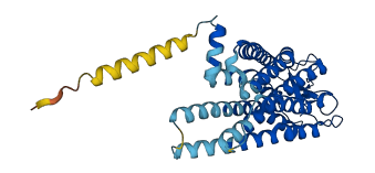
Descriptions
The autoinhibited protein was predicted that may have potential autoinhibitory elements via cis-regPred.
Autoinhibitory domains (AIDs)
Target domain |
|
Relief mechanism |
|
Assay |
cis-regPred |
Accessory elements
No accessory elements
Autoinhibited structure

Activated structure
1 structures for P79863
| Entry ID | Method | Resolution | Chain | Position | Source |
|---|---|---|---|---|---|
| AF-P79863-F1 | Predicted | AlphaFoldDB |
No variants for P79863
| Variant ID(s) | Position | Change | Description | Diseaes Association | Provenance |
|---|---|---|---|---|---|
| No variants for P79863 | |||||
No associated diseases with P79863
Functions
3 GO annotations of cellular component
| Name | Definition |
|---|---|
| integral component of membrane | The component of a membrane consisting of the gene products and protein complexes having at least some part of their peptide sequence embedded in the hydrophobic region of the membrane. |
| photoreceptor disc membrane | Stack of disc membranes located inside a photoreceptor outer segment, and containing densely packed molecules of photoreceptor proteins that traverse the lipid bilayer. Disc membranes arise as evaginations of the ciliary membrane during the development of the outer segment and may or may not remain contiguous with the ciliary membrane. |
| plasma membrane | The membrane surrounding a cell that separates the cell from its external environment. It consists of a phospholipid bilayer and associated proteins. |
2 GO annotations of molecular function
| Name | Definition |
|---|---|
| 11-cis retinal binding | Binding to 11-cis retinal, an isomer of retinal that plays an important role in the visual process in most vertebrates. 11-cis retinal combines with opsin in the rods (scotopsin) to form rhodopsin or visual purple. Retinal is one of the three compounds that makes up vitamin A. |
| G protein-coupled photoreceptor activity | Combining with incidental electromagnetic radiation, particularly visible light, and transmitting the signal across the membrane by activating an associated G-protein; promotes the exchange of GDP for GTP on the alpha subunit of a heterotrimeric G-protein complex. |
3 GO annotations of biological process
| Name | Definition |
|---|---|
| absorption of visible light | The reception of a (visible light) photon by a cell, visible light being defined as having a wavelength within the range 380-780 nm. |
| rhodopsin mediated signaling pathway | A G protein-coupled receptor signaling pathway initiated by the excitation of rhodopsin by a photon, and ending with the regulation of a downstream cellular process. |
| visual perception | The series of events required for an organism to receive a visual stimulus, convert it to a molecular signal, and recognize and characterize the signal. Visual stimuli are detected in the form of photons and are processed to form an image. |
No homologous proteins in AiPD
| UniProt AC | Gene Name | Protein Name | Species | Evidence Code |
|---|---|---|---|---|
| No homologous proteins | ||||
| 10 | 20 | 30 | 40 | 50 | 60 |
| MNGTEGENFY | VPMSNKTGVV | RSPFDYPQYY | LGEPWMFSAL | AAYMFFLILT | GLPVNFLTLF |
| 70 | 80 | 90 | 100 | 110 | 120 |
| VTIQHKKLRQ | PLNYILLNLA | VSDLFMVFGG | FTTTIITSMN | GYFIFGPAGC | NFEGFFATLG |
| 130 | 140 | 150 | 160 | 170 | 180 |
| GEVGLWCLVV | LAIERYMVVC | KPMANFRFGS | QHAIIGVVFT | WIMALSCAGP | PLVGWSRYIP |
| 190 | 200 | 210 | 220 | 230 | 240 |
| EGLQCSCGVD | YYTMKPEVNN | ESFVIYMFVV | HFTIPLIVIF | FCYGRLVCTV | KEAAAQQQES |
| 250 | 260 | 270 | 280 | 290 | 300 |
| ESTQRAEREV | TRMVIIMVVA | FLICWVPYAS | VAFYIFINQG | CDFTPFFMTV | PAFFAKSSAV |
| 310 | 320 | 330 | 340 | 350 | |
| YNPLIYILMN | KQFRNCMITT | ICLGKNPFEE | EESTSASASK | TEASSVSSSQ | VAPA |