P56581
Gene name |
Tdg |
Protein name |
G/T mismatch-specific thymine DNA glycosylase |
Names |
C-JUN leucine zipper interactive protein JZA-3, Thymine-DNA glycosylase |
Species |
Mus musculus (Mouse) |
KEGG Pathway |
mmu:21665 |
EC number |
3.2.2.29: Hydrolyzing N-glycosyl compounds |
Protein Class |
|
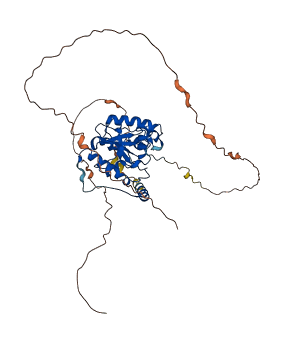
Descriptions
The autoinhibited protein was predicted that may have potential autoinhibitory elements via cis-regPred.
Autoinhibitory domains (AIDs)
Target domain |
|
Relief mechanism |
|
Assay |
cis-regPred |
Accessory elements
No accessory elements
Autoinhibited structure

Activated structure
1 structures for P56581
| Entry ID | Method | Resolution | Chain | Position | Source |
|---|---|---|---|---|---|
| AF-P56581-F1 | Predicted | AlphaFoldDB |
30 variants for P56581
| Variant ID(s) | Position | Change | Description | Diseaes Association | Provenance |
|---|---|---|---|---|---|
| rs49510163 | 57 | E>A | No | EVA | |
| rs219595175 | 57 | E>K | No | EVA | |
| rs3389124579 | 105 | K>R | No | EVA | |
| rs3389120786 | 117 | R>G | No | EVA | |
| rs3389084465 | 122 | F>Y | No | EVA | |
| rs3389120774 | 155 | M>T | No | EVA | |
| rs3389095386 | 167 | G>A | No | EVA | |
| rs3389059472 | 167 | G>R | No | EVA | |
| rs3401644503 | 179 | L>A | No | EVA | |
| rs243823394 | 181 | E>D | No | EVA | |
| rs3389084387 | 183 | Q>* | No | EVA | |
| rs3389117571 | 187 | M>I | No | EVA | |
| rs3389118007 | 211 | S>N | No | EVA | |
| rs3389120555 | 226 | L>M | No | EVA | |
| rs3389113693 | 250 | S>N | No | EVA | |
| rs3389105689 | 277 | C>W | No | EVA | |
| rs3389128222 | 281 | P>S | No | EVA | |
| rs3389120782 | 288 | A>T | No | EVA | |
| rs3389092760 | 291 | P>S | No | EVA | |
| rs47249452 | 317 | A>S | No | EVA | |
| rs47249452 | 317 | A>T | No | EVA | |
| rs248551590 | 319 | V>I | No | EVA | |
| rs3389105671 | 338 | M>K | No | EVA | |
| rs3389117536 | 344 | K>N | No | EVA | |
| rs3401000586 | 378 | S>R | No | EVA | |
| rs229431027 | 379 | A>S | No | EVA | |
| rs47946519 | 386 | A>T | No | EVA | |
| rs47182293 | 388 | S>G | No | EVA | |
| rs581922266 | 414 | E>D | No | EVA | |
| rs585881083 | 421 | A>T | No | EVA |
No associated diseases with P56581
1 regional properties for P56581
| Type | Name | Position | InterPro Accession |
|---|---|---|---|
| domain | Uracil-DNA glycosylase-like | 142 - 291 | IPR005122 |
Functions
| Description | ||
|---|---|---|
| EC Number | 3.2.2.29 | Hydrolyzing N-glycosyl compounds |
| Subcellular Localization |
|
|
| PANTHER Family | ||
| PANTHER Subfamily | ||
| PANTHER Protein Class | ||
| PANTHER Pathway Category | No pathway information available | |
4 GO annotations of cellular component
| Name | Definition |
|---|---|
| nucleoplasm | That part of the nuclear content other than the chromosomes or the nucleolus. |
| nucleus | A membrane-bounded organelle of eukaryotic cells in which chromosomes are housed and replicated. In most cells, the nucleus contains all of the cell's chromosomes except the organellar chromosomes, and is the site of RNA synthesis and processing. In some species, or in specialized cell types, RNA metabolism or DNA replication may be absent. |
| plasma membrane | The membrane surrounding a cell that separates the cell from its external environment. It consists of a phospholipid bilayer and associated proteins. |
| PML body | A class of nuclear body; they react against SP100 auto-antibodies (PML, promyelocytic leukemia); cells typically contain 10-30 PML bodies per nucleus; alterations in the localization of PML bodies occurs after viral infection. |
18 GO annotations of molecular function
| Name | Definition |
|---|---|
| ATP binding | Binding to ATP, adenosine 5'-triphosphate, a universally important coenzyme and enzyme regulator. |
| chloride ion binding | Binding to a chloride ion (Cl-). |
| DNA binding | Any molecular function by which a gene product interacts selectively and non-covalently with DNA (deoxyribonucleic acid). |
| DNA N-glycosylase activity | Catalysis of the removal of damaged bases by cleaving the N-C1' glycosidic bond between the target damaged DNA base and the deoxyribose sugar. The reaction releases a free base and leaves an apurinic/apyrimidinic (AP) site. |
| DNA-binding transcription factor binding | Binding to a DNA-binding transcription factor, a protein that interacts with a specific DNA sequence (sometimes referred to as a motif) within the regulatory region of a gene to modulate transcription. |
| double-stranded DNA binding | Binding to double-stranded DNA. |
| G/U mismatch-specific uracil-DNA glycosylase activity | Catalysis of the removal of uracil from a U*G mispair by the cleavage the N-C1' glycosidic bond between the target damaged DNA base and the deoxyribose sugar. The reaction releases a free uracil and leaves an apyrimidinic (AP) site. |
| magnesium ion binding | Binding to a magnesium (Mg) ion. |
| mismatched DNA binding | Binding to a double-stranded DNA region containing one or more mismatches. |
| protein domain specific binding | Binding to a specific domain of a protein. |
| protein homodimerization activity | Binding to an identical protein to form a homodimer. |
| protein kinase C binding | Binding to protein kinase C. |
| protein self-association | Binding to a domain within the same polypeptide. |
| pyrimidine-specific mismatch base pair DNA N-glycosylase activity | Catalysis of the removal of mismatched pyrimidine bases in DNA. Enzymes with this activity recognize and remove pyrimidines present in mismatches by cleaving the N-C1' glycosidic bond between the target damaged DNA base and the deoxyribose sugar. The reaction releases a free base and leaves an apyrimidinic (AP) site. |
| sodium ion binding | Binding to a sodium ion (Na+). |
| SUMO binding | Binding to the small ubiquitin-like protein SUMO. |
| transcription coregulator activity | A transcription regulator activity that modulates the transcription of specific gene sets via binding to a DNA-bound DNA-binding transcription factor, either on its own or as part of a complex. Coregulators often act by altering chromatin structure and modifications. For example, one class of transcription coregulators modifies chromatin structure through covalent modification of histones. A second class remodels the conformation of chromatin in an ATP-dependent fashion. A third class modulates interactions of DNA-bound DNA-binding transcription factors with other transcription coregulators. |
| uracil DNA N-glycosylase activity | Catalysis of the cleavage of the N-C1' glycosidic bond between the damaged DNA base and the deoxyribose sugar, releasing a free base and leaving an apyrimidinic (AP) site. Enzymes with this activity recognize and remove uracil bases in DNA that result from the deamination of cytosine or the misincorporation of dUTP opposite an adenine. |
10 GO annotations of biological process
| Name | Definition |
|---|---|
| base-excision repair | In base excision repair, an altered base is removed by a DNA glycosylase enzyme, followed by excision of the resulting sugar phosphate. The small gap left in the DNA helix is filled in by the sequential action of DNA polymerase and DNA ligase. |
| base-excision repair, AP site formation | The formation of an AP site, a deoxyribose sugar with a missing base, by DNA glycosylase which recognizes an altered base in DNA and catalyzes its hydrolytic removal. This sugar phosphate is the substrate recognized by the AP endonuclease, which cuts the DNA phosphodiester backbone at the 5' side of the altered site to leave a gap which is subsequently repaired. |
| DNA demethylation | The removal of a methyl group from one or more nucleotides within an DNA molecule. |
| mismatch repair | A system for the correction of errors in which an incorrect base, which cannot form hydrogen bonds with the corresponding base in the parent strand, is incorporated into the daughter strand. The mismatch repair system promotes genomic fidelity by repairing base-base mismatches, insertion-deletion loops and heterologies generated during DNA replication and recombination. |
| negative regulation of chromatin binding | Any process that stops or reduces the frequency, rate or extent of chromatin binding. Chromatin binding is the selective interaction with chromatin, the network of fibers of DNA, protein, and sometimes RNA, that make up the chromosomes of the eukaryotic nucleus during interphase. |
| negative regulation of protein binding | Any process that stops, prevents, or reduces the frequency, rate or extent of protein binding. |
| negative regulation of transcription by RNA polymerase II | Any process that stops, prevents, or reduces the frequency, rate or extent of transcription mediated by RNA polymerase II. |
| regulation of DNA N-glycosylase activity | Any process that modulates the frequency, rate or extent of DNA N-glycosylase activity. |
| regulation of embryonic development | Any process that modulates the frequency, rate or extent of embryonic development. |
| regulation of gene expression, epigenetic | A process that modulates the frequency, rate or extent of gene expression through chromatin remodelling either by modifying higher order chromatin fiber structure, nucleosomal histones, or the DNA. Once established, this regulation may be maintained over many cell divisions. It can also be heritable in the absence of the instigating signal. |
No homologous proteins in AiPD
| UniProt AC | Gene Name | Protein Name | Species | Evidence Code |
|---|---|---|---|---|
| No homologous proteins | ||||
| 10 | 20 | 30 | 40 | 50 | 60 |
| MDAEAARSYS | LEQVQALYSF | PFQQMMAEVP | NMAVTTGQQV | PAVAPNMATV | TEQQVPEDAP |
| 70 | 80 | 90 | 100 | 110 | 120 |
| VQEPAPEAPK | RRKRKPRAAE | PQEPVEPKKP | ATSKKSGKST | KSKEKQEKIT | DAFKVKRKVD |
| 130 | 140 | 150 | 160 | 170 | 180 |
| RFNGVSEAEL | LTKTLPDILT | FNLDIVIIGI | NPGLMAAYKG | HHYPGPGNHF | WKCLFMSGLS |
| 190 | 200 | 210 | 220 | 230 | 240 |
| EVQLNHMDDH | TLPGKYGIGF | TNMVERTTPG | SKDLSSKEFR | EGGRILVQKL | QKYQPRIAVF |
| 250 | 260 | 270 | 280 | 290 | 300 |
| NGKCIYEIFS | KEVFGVKVKN | LEFGLQPHKI | PDTETLCYVM | PSSSARCAQF | PRAQDKVHYY |
| 310 | 320 | 330 | 340 | 350 | 360 |
| IKLKDLRDQL | KGIERNADVQ | EVQYTFDLQL | AQEDAKKMAV | KEEKYDPGYE | AAYGGAYGEN |
| 370 | 380 | 390 | 400 | 410 | 420 |
| PCNGEPCGIA | SNGLTAHSAE | PRGEAAPSDV | PNGQWMAQSF | AEQIPSFNNC | GTREQEEESH |
| A |