P54680
Gene name |
fimA (DDB_G0277855) |
Protein name |
Fimbrin |
Names |
|
Species |
Dictyostelium discoideum (Slime mold) |
KEGG Pathway |
ddi:DDB_G0277855 |
EC number |
|
Protein Class |
|
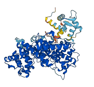
Descriptions
The autoinhibited protein was predicted that may have potential autoinhibitory elements via cis-regPred.
Autoinhibitory domains (AIDs)
Target domain |
|
Relief mechanism |
|
Assay |
cis-regPred |
Accessory elements
No accessory elements
Autoinhibited structure

Activated structure
1 structures for P54680
| Entry ID | Method | Resolution | Chain | Position | Source |
|---|---|---|---|---|---|
| AF-P54680-F1 | Predicted | AlphaFoldDB |
No variants for P54680
| Variant ID(s) | Position | Change | Description | Diseaes Association | Provenance |
|---|---|---|---|---|---|
| No variants for P54680 | |||||
No associated diseases with P54680
10 regional properties for P54680
| Type | Name | Position | InterPro Accession |
|---|---|---|---|
| conserved_site | Actinin-type actin-binding domain, conserved site | 204 - 228 | IPR001589-1 |
| conserved_site | Actinin-type actin-binding domain, conserved site | 381 - 390 | IPR001589-2 |
| conserved_site | Actinin-type actin-binding domain, conserved site | 460 - 484 | IPR001589-3 |
| domain | Calponin homology domain | 116 - 232 | IPR001715-1 |
| domain | Calponin homology domain | 260 - 365 | IPR001715-2 |
| domain | Calponin homology domain | 379 - 488 | IPR001715-3 |
| domain | Calponin homology domain | 501 - 608 | IPR001715-4 |
| domain | EF-hand domain | 7 - 78 | IPR002048 |
| binding_site | EF-Hand 1, calcium-binding site | 20 - 32 | IPR018247-1 |
| binding_site | EF-Hand 1, calcium-binding site | 56 - 68 | IPR018247-2 |
7 GO annotations of cellular component
| Name | Definition |
|---|---|
| actin filament | A filamentous structure formed of a two-stranded helical polymer of the protein actin and associated proteins. Actin filaments are a major component of the contractile apparatus of skeletal muscle and the microfilaments of the cytoskeleton of eukaryotic cells. The filaments, comprising polymerized globular actin molecules, appear as flexible structures with a diameter of 5-9 nm. They are organized into a variety of linear bundles, two-dimensional networks, and three dimensional gels. In the cytoskeleton they are most highly concentrated in the cortex of the cell just beneath the plasma membrane. |
| actin filament bundle | An assembly of actin filaments that are on the same axis but may be oriented with the same or opposite polarities and may be packed with different levels of tightness. |
| cell leading edge | The area of a motile cell closest to the direction of movement. |
| cytoplasm | The contents of a cell excluding the plasma membrane and nucleus, but including other subcellular structures. |
| lamellipodium | A thin sheetlike process extended by the leading edge of a migrating cell or extending cell process; contains a dense meshwork of actin filaments. |
| phagocytic cup | An invagination of the cell membrane formed by an actin dependent process during phagocytosis. Following internalization it is converted into a phagosome. |
| phagocytic vesicle | A membrane-bounded intracellular vesicle that arises from the ingestion of particulate material by phagocytosis. |
2 GO annotations of molecular function
| Name | Definition |
|---|---|
| actin filament binding | Binding to an actin filament, also known as F-actin, a helical filamentous polymer of globular G-actin subunits. |
| calcium ion binding | Binding to a calcium ion (Ca2+). |
3 GO annotations of biological process
| Name | Definition |
|---|---|
| actin filament bundle assembly | The assembly of actin filament bundles; actin filaments are on the same axis but may be oriented with the same or opposite polarities and may be packed with different levels of tightness. |
| actin filament network formation | The assembly of a network of actin filaments; actin filaments on different axes and with differing orientations are crosslinked together to form a mesh of filaments. |
| sexual reproduction | A type of reproduction that combines the genetic material of two gametes (such as a sperm or egg cell or fungal spores). The gametes have an haploid genome (with a single set of chromosomes, the product of a meiotic division) and combines with one another to produce a zygote (diploid). |
No homologous proteins in AiPD
| UniProt AC | Gene Name | Protein Name | Species | Evidence Code |
|---|---|---|---|---|
| No homologous proteins | ||||
| 10 | 20 | 30 | 40 | 50 | 60 |
| MTNFSESEIS | EFKASFNQFD | ENGDGQISAL | ELQKILTKCG | EKVTGVEVRD | MIKEVDTDGN |
| 70 | 80 | 90 | 100 | 110 | 120 |
| GSIDFKEFLQ | VMQKARQHSA | NASPAFASAV | KKVGAVNTIG | GYSGSTASGV | QHSYSDEEKV |
| 130 | 140 | 150 | 160 | 170 | 180 |
| AYIDWINNCL | AKDVDLKSRL | PIPEDGDKFF | AACNDGLLLC | KLINDAVPDT | IDERVLNKKN |
| 190 | 200 | 210 | 220 | 230 | 240 |
| LNAFRINENQ | VLCINSAKAI | GCNVVNIGAG | DLVEGRAHLI | MGLTWQIIKI | GLFARINLTN |
| 250 | 260 | 270 | 280 | 290 | 300 |
| HPELYRLLHD | GETIEDLLKL | PVEEILLRWF | NYHLAAAGSQ | RRVKNFSGDI | KDSECYTILL |
| 310 | 320 | 330 | 340 | 350 | 360 |
| KQIAPKDAGV | ETSALNISNL | DQRAVKVLEN | ADKLGCKKFL | KPKDIVTGFQ | KLNLAFVANL |
| 370 | 380 | 390 | 400 | 410 | 420 |
| FNTHPALEPV | EDVVIIEETR | EEKTFRNWMN | SLGVDPFVNN | LYEGTYDGLI | LIQLFDKIYP |
| 430 | 440 | 450 | 460 | 470 | 480 |
| GLVDHKKVNY | PPYKAMGAEM | KKIENCNYAI | QLGKDCKYSL | VGIDGKNVYD | KNKTLTLSIL |
| 490 | 500 | 510 | 520 | 530 | 540 |
| WQLMRGHVIS | ILTALSGSGK | PIADADIVNW | TNSKLSAAGK | KQISGFKDST | ISTGIPILDV |
| 550 | 560 | 570 | 580 | 590 | 600 |
| IEAVRPGSVD | PALVATSGSA | EDNLLNAKLA | VSTARKVGAV | VFALPEDIVE | VKPKMVLTLF |
| ASLWQVEMTK |