P41592
Gene name |
|
Protein name |
Red-sensitive opsin |
Names |
Red cone photoreceptor pigment |
Species |
Anolis carolinensis (Green anole) (American chameleon) |
KEGG Pathway |
acs:100564371 |
EC number |
|
Protein Class |
|
Descriptions
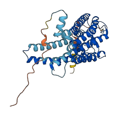
The autoinhibited protein was predicted that may have potential autoinhibitory elements via cis-regPred.
Autoinhibitory domains (AIDs)
Target domain |
|
Relief mechanism |
|
Assay |
cis-regPred |
Accessory elements
No accessory elements
Autoinhibited structure

Activated structure
1 structures for P41592
| Entry ID | Method | Resolution | Chain | Position | Source |
|---|---|---|---|---|---|
| AF-P41592-F1 | Predicted | AlphaFoldDB |
No variants for P41592
| Variant ID(s) | Position | Change | Description | Diseaes Association | Provenance |
|---|---|---|---|---|---|
| No variants for P41592 | |||||
No associated diseases with P41592
5 GO annotations of cellular component
| Name | Definition |
|---|---|
| cell body | The portion of a cell bearing surface projections such as axons, dendrites, cilia, or flagella that includes the nucleus, but excludes all cell projections. |
| integral component of plasma membrane | The component of the plasma membrane consisting of the gene products and protein complexes having at least some part of their peptide sequence embedded in the hydrophobic region of the membrane. |
| photoreceptor inner segment membrane | The membrane surrounding the inner segment of a vertebrate photoreceptor. The photoreceptor inner segment contains mitochondria, ribosomes and membranes where opsin molecules are assembled and passed to be part of the outer segment discs. |
| photoreceptor outer segment | The outer segment of a vertebrate photoreceptor that contains a stack of membrane discs embedded with photoreceptor proteins. |
| vesicle | Any small, fluid-filled, spherical organelle enclosed by membrane. |
3 GO annotations of molecular function
| Name | Definition |
|---|---|
| chloride ion binding | Binding to a chloride ion (Cl-). |
| G protein-coupled photoreceptor activity | Combining with incidental electromagnetic radiation, particularly visible light, and transmitting the signal across the membrane by activating an associated G-protein; promotes the exchange of GDP for GTP on the alpha subunit of a heterotrimeric G-protein complex. |
| protein homodimerization activity | Binding to an identical protein to form a homodimer. |
7 GO annotations of biological process
| Name | Definition |
|---|---|
| absorption of visible light | The reception of a (visible light) photon by a cell, visible light being defined as having a wavelength within the range 380-780 nm. |
| cellular response to light stimulus | Any process that results in a change in state or activity of a cell (in terms of movement, secretion, enzyme production, gene expression, etc.) as a result of a light stimulus, electromagnetic radiation of wavelengths classified as infrared, visible or ultraviolet light. |
| G protein-coupled receptor signaling pathway | The series of molecular signals initiated by a ligand binding to its receptor, in which the activated receptor promotes the exchange of GDP for GTP on the alpha-subunit of an associated heterotrimeric G-protein complex. The GTP-bound activated alpha-G-protein then dissociates from the beta- and gamma-subunits to further transmit the signal within the cell. The pathway begins with receptor-ligand interaction, and ends with regulation of a downstream cellular process. The pathway can start from the plasma membrane, Golgi or nuclear membrane. |
| light absorption | The reception of a photon by a cell. |
| phototransduction | The sequence of reactions within a cell required to convert absorbed photons into a molecular signal. |
| protein homotrimerization | The formation of a protein homotrimer, a macromolecular structure consisting of three noncovalently associated identical subunits. |
| visual perception | The series of events required for an organism to receive a visual stimulus, convert it to a molecular signal, and recognize and characterize the signal. Visual stimuli are detected in the form of photons and are processed to form an image. |
No homologous proteins in AiPD
| UniProt AC | Gene Name | Protein Name | Species | Evidence Code |
|---|---|---|---|---|
| No homologous proteins | ||||
| 10 | 20 | 30 | 40 | 50 | 60 |
| MAGTVTEAWD | VAVFAARRRN | DEDDTTRDSL | FTYTNSNNTR | GPFEGPNYHI | APRWVYNITS |
| 70 | 80 | 90 | 100 | 110 | 120 |
| VWMIFVVIAS | IFTNGLVLVA | TAKFKKLRHP | LNWILVNLAI | ADLGETVIAS | TISVINQISG |
| 130 | 140 | 150 | 160 | 170 | 180 |
| YFILGHPMCV | LEGYTVSTCG | ISALWSLAVI | SWERWVVVCK | PFGNVKFDAK | LAVAGIVFSW |
| 190 | 200 | 210 | 220 | 230 | 240 |
| VWSAVWTAPP | VFGWSRYWPH | GLKTSCGPDV | FSGSDDPGVL | SYMIVLMITC | CFIPLAVILL |
| 250 | 260 | 270 | 280 | 290 | 300 |
| CYLQVWLAIR | AVAAQQKESE | STQKAEKEVS | RMVVVMIIAY | CFCWGPYTVF | ACFAAANPGY |
| 310 | 320 | 330 | 340 | 350 | 360 |
| AFHPLAAALP | AYFAKSATIY | NPIIYVFMNR | QFRNCIMQLF | GKKVDDGSEL | SSTSRTEVSS |
| VSNSSVSPA |