P38410
Gene name |
gnaq |
Protein name |
Guanine nucleotide-binding protein G(q) subunit alpha |
Names |
Guanine nucleotide-binding protein alpha-q |
Species |
Xenopus laevis (African clawed frog) |
KEGG Pathway |
xla:399261 |
EC number |
|
Protein Class |
|
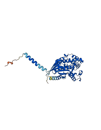
Descriptions
The autoinhibited protein was predicted that may have potential autoinhibitory elements via cis-regPred.
Autoinhibitory domains (AIDs)
Target domain |
|
Relief mechanism |
|
Assay |
cis-regPred |
Accessory elements
No accessory elements
Autoinhibited structure

Activated structure
1 structures for P38410
| Entry ID | Method | Resolution | Chain | Position | Source |
|---|---|---|---|---|---|
| AF-P38410-F1 | Predicted | AlphaFoldDB |
No variants for P38410
| Variant ID(s) | Position | Change | Description | Diseaes Association | Provenance |
|---|---|---|---|---|---|
| No variants for P38410 | |||||
No associated diseases with P38410
No regional properties for P38410
| Type | Name | Position | InterPro Accession |
|---|---|---|---|
| No domain, repeats, and functional sites for P38410 | |||
6 GO annotations of cellular component
| Name | Definition |
|---|---|
| Golgi apparatus | A membrane-bound cytoplasmic organelle of the endomembrane system that further processes the core oligosaccharides (e.g. N-glycans) added to proteins in the endoplasmic reticulum and packages them into membrane-bound vesicles. The Golgi apparatus operates at the intersection of the secretory, lysosomal, and endocytic pathways. |
| membrane | A lipid bilayer along with all the proteins and protein complexes embedded in it an attached to it. |
| nuclear membrane | Either of the lipid bilayers that surround the nucleus and form the nuclear envelope; excludes the intermembrane space. |
| photoreceptor outer segment | The outer segment of a vertebrate photoreceptor that contains a stack of membrane discs embedded with photoreceptor proteins. |
| plasma membrane | The membrane surrounding a cell that separates the cell from its external environment. It consists of a phospholipid bilayer and associated proteins. |
| synapse | The junction between an axon of one neuron and a dendrite of another neuron, a muscle fiber or a glial cell. As the axon approaches the synapse it enlarges into a specialized structure, the presynaptic terminal bouton, which contains mitochondria and synaptic vesicles. At the tip of the terminal bouton is the presynaptic membrane; facing it, and separated from it by a minute cleft (the synaptic cleft) is a specialized area of membrane on the receiving cell, known as the postsynaptic membrane. In response to the arrival of nerve impulses, the presynaptic terminal bouton secretes molecules of neurotransmitters into the synaptic cleft. These diffuse across the cleft and transmit the signal to the postsynaptic membrane. |
5 GO annotations of molecular function
| Name | Definition |
|---|---|
| G protein-coupled receptor binding | Binding to a G protein-coupled receptor. |
| G-protein beta/gamma-subunit complex binding | Binding to a complex of G-protein beta/gamma subunits. |
| GTP binding | Binding to GTP, guanosine triphosphate. |
| GTPase activity | Catalysis of the reaction: GTP + H2O = GDP + H+ + phosphate. |
| metal ion binding | Binding to a metal ion. |
3 GO annotations of biological process
| Name | Definition |
|---|---|
| entrainment of circadian clock | The synchronization of a circadian rhythm to environmental time cues such as light. |
| G protein-coupled acetylcholine receptor signaling pathway | A G protein-coupled receptor signaling pathway initiated by a ligand binding to an acetylcholine receptor on the surface of a target cell, and ends with regulation of a downstream cellular process, e.g. transcription. |
| phototransduction, visible light | The sequence of reactions within a cell required to convert absorbed photons from visible light into a molecular signal. A visible light stimulus is electromagnetic radiation that can be perceived visually by an organism; for organisms lacking a visual system, this can be defined as light with a wavelength within the range 380 to 780 nm. |
No homologous proteins in AiPD
| UniProt AC | Gene Name | Protein Name | Species | Evidence Code |
|---|---|---|---|---|
| No homologous proteins | ||||
| 10 | 20 | 30 | 40 | 50 | 60 |
| MTLESIMACC | LSEEAEEARR | INDEIERQLR | RDKRDARREL | KLLLLGTGES | GKSTFIKQMR |
| 70 | 80 | 90 | 100 | 110 | 120 |
| IIHGSGYSDE | DKRGFTKLVY | QNIFSAMQAM | IRAMETLKIP | YKYEHNKGHA | LLVREVDVEK |
| 130 | 140 | 150 | 160 | 170 | 180 |
| VASFENPYVD | AIKYLWNDPG | IQECYDRRRE | YQLSDSTKYY | LNDLDRIATH | GYLPTQQDVL |
| 190 | 200 | 210 | 220 | 230 | 240 |
| RVRVPTTGII | EYPFDLQSVI | FRMVDVGGQR | SERRKWIHCF | ENVTSIMFLV | ALSEYDQVLV |
| 250 | 260 | 270 | 280 | 290 | 300 |
| ESDNENRMEE | SKALFRTIIT | YPWFQNSSVI | LFLNKKDLLE | EKIMYSHLVD | YFPEYDGPQR |
| 310 | 320 | 330 | 340 | 350 | |
| DAQAAREFIL | KMFVDLNPDS | DKIIYSHFTC | ATDTENIRFV | FAAVKDTILQ | LNLKEYNLV |