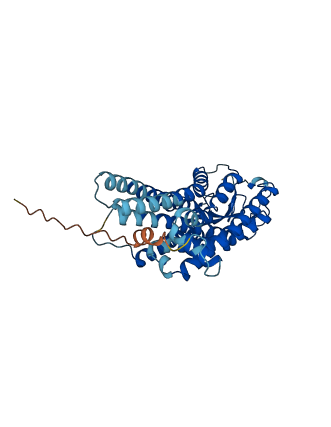
Descriptions
The autoinhibited protein was predicted that may have potential autoinhibitory elements via cis-regPred.
Autoinhibitory domains (AIDs)
Target domain |
|
Relief mechanism |
|
Assay |
cis-regPred |
Accessory elements
No accessory elements
Autoinhibited structure

Activated structure
1 structures for P35357
| Entry ID | Method | Resolution | Chain | Position | Source |
|---|---|---|---|---|---|
| AF-P35357-F1 | Predicted | AlphaFoldDB |
No variants for P35357
| Variant ID(s) | Position | Change | Description | Diseaes Association | Provenance |
|---|---|---|---|---|---|
| No variants for P35357 | |||||
No associated diseases with P35357
1 GO annotations of cellular component
| Name | Definition |
|---|---|
| integral component of membrane | The component of a membrane consisting of the gene products and protein complexes having at least some part of their peptide sequence embedded in the hydrophobic region of the membrane. |
2 GO annotations of molecular function
| Name | Definition |
|---|---|
| G protein-coupled receptor activity | Combining with an extracellular signal and transmitting the signal across the membrane by activating an associated G-protein; promotes the exchange of GDP for GTP on the alpha subunit of a heterotrimeric G-protein complex. |
| photoreceptor activity | The function of absorbing and responding to incidental electromagnetic radiation, particularly visible light. The response may involve a change in conformation. |
2 GO annotations of biological process
| Name | Definition |
|---|---|
| phototransduction | The sequence of reactions within a cell required to convert absorbed photons into a molecular signal. |
| visual perception | The series of events required for an organism to receive a visual stimulus, convert it to a molecular signal, and recognize and characterize the signal. Visual stimuli are detected in the form of photons and are processed to form an image. |
No homologous proteins in AiPD
| UniProt AC | Gene Name | Protein Name | Species | Evidence Code |
|---|---|---|---|---|
| No homologous proteins | ||||
| 10 | 20 | 30 | 40 | 50 | 60 |
| MNGTEGINFY | VPLSNKTGLV | RSPFEYPQYY | LADPWKFKVL | SFYMFFLIAA | GMPLNGLTLF |
| 70 | 80 | 90 | 100 | 110 | 120 |
| VTFQHKKLRQ | PLNYILVNLA | AANLVTVCCG | FTVTFYASWY | AYFVFGPIGC | AIEGFFATIG |
| 130 | 140 | 150 | 160 | 170 | 180 |
| GQVALWSLVV | LAIERYIVIC | KPMGNFRFSA | THAIMGIAFT | WFMALACAGP | PLFGWSRFIP |
| 190 | 200 | 210 | 220 | 230 | 240 |
| EGMQCSCGPD | YYTLNPDFHN | ESYVIYMFIV | HFTVPMVVIF | FSYGRLVCKV | REAAAQQQES |
| 250 | 260 | 270 | 280 | 290 | 300 |
| ATTQKAEKEV | TRMVILMVLG | FLLAWTPYAA | TAIWIFTNRG | AAFSVTFMTI | PAFFSKSSSI |
| 310 | 320 | 330 | 340 | 350 | |
| YNPIIYVLLN | KQFRNCMVTT | ICCGKNPFGD | EDVSSSVSQS | KTEVSSVSSS | QVAPA |