P11966
Gene name |
PDHB |
Protein name |
Pyruvate dehydrogenase E1 component subunit beta, mitochondrial |
Names |
PDHE1-B |
Species |
Bos taurus (Bovine) |
KEGG Pathway |
bta:613610 |
EC number |
1.2.4.1: With a disulfide as acceptor |
Protein Class |
|
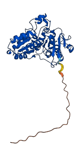
Descriptions
The autoinhibited protein was predicted that may have potential autoinhibitory elements via cis-regPred.
Autoinhibitory domains (AIDs)
Target domain |
|
Relief mechanism |
|
Assay |
cis-regPred |
Accessory elements
No accessory elements
Autoinhibited structure

Activated structure
1 structures for P11966
| Entry ID | Method | Resolution | Chain | Position | Source |
|---|---|---|---|---|---|
| AF-P11966-F1 | Predicted | AlphaFoldDB |
38 variants for P11966
| Variant ID(s) | Position | Change | Description | Diseaes Association | Provenance |
|---|---|---|---|---|---|
| rs457497421 | 2 | A>G | No | EVA | |
| rs442854960 | 3 | V>A | No | EVA | |
| rs442854960 | 3 | V>G | No | EVA | |
| rs475912647 | 3 | V>L | No | EVA | |
| rs133136297 | 4 | V>A | No | EVA | |
| rs133136297 | 4 | V>G | No | EVA | |
| rs473384482 | 10 | K>N | No | EVA | |
| rs134725284 | 23 | F>V | No | EVA | |
| rs443261115 | 81 | R>G | No | EVA | |
| rs461719101 | 81 | R>S | No | EVA | |
| rs481779075 | 82 | I>V | No | EVA | |
| rs478999754 | 129 | K>N | No | EVA | |
| rs446134642 | 149 | G>A | No | EVA | |
| rs464518124 | 161 | C>F | No | EVA | |
| rs438020735 | 162 | F>L | No | EVA | |
| rs450011898 | 162 | F>Y | No | EVA | |
| rs435525613 | 166 | Y>S | No | EVA | |
| rs453988764 | 168 | H>Y | No | EVA | |
| rs472415003 | 169 | C>W | No | EVA | |
| rs432923832 | 170 | P>T | No | EVA | |
| rs451490346 | 171 | G>V | No | EVA | |
| rs476315160 | 173 | K>T | No | EVA | |
| rs443299891 | 177 | P>T | No | EVA | |
| rs455293844 | 180 | S>A | No | EVA | |
| rs473818972 | 182 | D>G | No | EVA | |
| rs460832826 | 190 | A>P | No | EVA | |
| rs479087369 | 192 | R>Q | No | EVA | |
| rs439775832 | 193 | D>Y | No | EVA | |
| rs458136323 | 194 | N>S | No | EVA | |
| rs482926586 | 195 | N>I | No | EVA | |
| rs468590319 | 197 | V>L | No | EVA | |
| rs134377717 | 200 | L>V | No | EVA | |
| rs441193887 | 233 | Q>* | No | EVA | |
| rs454726519 | 289 | L>I | No | EVA | |
| rs471749860 | 319 | D>N | No | EVA | |
| rs451046654 | 330 | V>G | No | EVA | |
| rs469472806 | 340 | D>V | No | EVA | |
| rs436450654 | 346 | V>F | No | EVA |
No associated diseases with P11966
Functions
| Description | ||
|---|---|---|
| EC Number | 1.2.4.1 | With a disulfide as acceptor |
| Subcellular Localization |
|
|
| PANTHER Family | ||
| PANTHER Subfamily | ||
| PANTHER Protein Class | ||
| PANTHER Pathway Category | No pathway information available | |
2 GO annotations of cellular component
| Name | Definition |
|---|---|
| mitochondrial pyruvate dehydrogenase complex | Complex that carries out the oxidative decarboxylation of pyruvate to form acetyl-CoA in eukaryotes; includes subunits possessing three catalytic activities: pyruvate dehydrogenase (E1), dihydrolipoamide S-acetyltransferase (E2), and dihydrolipoamide dehydrogenase (E3). The This Eukaryotic form usually contains more subunits than its bacterial counterpart; for example, one known complex contains 30 E1 dimers, 60 E2 monomers, and 6 E3 dimers as well as a few copies of pyruvate dehydrogenase kinase and pyruvate dehydrogenase phosphatase. |
| pyruvate dehydrogenase complex | Complex that carries out the oxidative decarboxylation of pyruvate to form acetyl-CoA; comprises subunits possessing three catalytic activities: pyruvate dehydrogenase (E1), dihydrolipoamide S-acetyltransferase (E2), and dihydrolipoamide dehydrogenase (E3). |
1 GO annotations of molecular function
| Name | Definition |
|---|---|
| pyruvate dehydrogenase (acetyl-transferring) activity | Catalysis of the reaction: pyruvate + lipoamide = S-acetyldihydrolipoamide + CO2. |
3 GO annotations of biological process
| Name | Definition |
|---|---|
| acetyl-CoA biosynthetic process from pyruvate | The chemical reactions and pathways resulting in the formation of acetyl-CoA from pyruvate. |
| glucose metabolic process | The chemical reactions and pathways involving glucose, the aldohexose gluco-hexose. D-glucose is dextrorotatory and is sometimes known as dextrose; it is an important source of energy for living organisms and is found free as well as combined in homo- and hetero-oligosaccharides and polysaccharides. |
| tricarboxylic acid cycle | A nearly universal metabolic pathway in which the acetyl group of acetyl coenzyme A is effectively oxidized to two CO2 and four pairs of electrons are transferred to coenzymes. The acetyl group combines with oxaloacetate to form citrate, which undergoes successive transformations to isocitrate, 2-oxoglutarate, succinyl-CoA, succinate, fumarate, malate, and oxaloacetate again, thus completing the cycle. In eukaryotes the tricarboxylic acid is confined to the mitochondria. See also glyoxylate cycle. |
3 homologous proteins in AiPD
| UniProt AC | Gene Name | Protein Name | Species | Evidence Code |
|---|---|---|---|---|
| P11177 | PDHB | Pyruvate dehydrogenase E1 component subunit beta, mitochondrial | Homo sapiens (Human) | PR |
| Q9D051 | Pdhb | Pyruvate dehydrogenase E1 component subunit beta, mitochondrial | Mus musculus (Mouse) | PR |
| P49432 | Pdhb | Pyruvate dehydrogenase E1 component subunit beta, mitochondrial | Rattus norvegicus (Rat) | PR |
| 10 | 20 | 30 | 40 | 50 | 60 |
| MAVVAVLVRK | PLEQVSGLLR | RRFHRTAPAA | LQVTVREAIN | QGMDEELERD | EKVFLLGEEV |
| 70 | 80 | 90 | 100 | 110 | 120 |
| AQYDGAYKVS | RGLWKKYGDK | RIIDTPISEM | GFAGIAVGAA | MAGLRPICEF | MTFNFSMQAI |
| 130 | 140 | 150 | 160 | 170 | 180 |
| DQVINSAAKT | YYMSGGLQSV | PIVFRGPNGA | SAGVAAQHSQ | CFAAWYGHCP | GLKVVSPWSS |
| 190 | 200 | 210 | 220 | 230 | 240 |
| EDAKGLIKSA | IRDNNPVVVL | ENELMYGVPF | ELPSEAQSKD | FLIPIGKAKI | ERQGTHVTIV |
| 250 | 260 | 270 | 280 | 290 | 300 |
| AHSRPVGHCL | EAATVLSKEG | IECEVINLRT | IRPMDIETIE | GSVMKTNHLV | TVEGGWPQFG |
| 310 | 320 | 330 | 340 | 350 | |
| VGAEICARIM | EGPAFNFLDA | PAVRVTGADV | PMPYAKILED | NSVPQVKDII | FAIKKTLNI |