P0AFG8
Gene name |
aceE (b0114, JW0110) |
Protein name |
Pyruvate dehydrogenase E1 component |
Names |
PDH E1 component |
Species |
Escherichia coli (strain K12) |
KEGG Pathway |
eco:b0114 |
EC number |
1.2.4.1: With a disulfide as acceptor |
Protein Class |
|
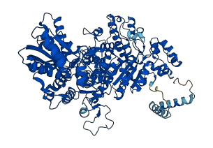
Descriptions
The autoinhibited protein was predicted that may have potential autoinhibitory elements via cis-regPred.
Autoinhibitory domains (AIDs)
Target domain |
|
Relief mechanism |
|
Assay |
cis-regPred |
Accessory elements
No accessory elements
Autoinhibited structure

Activated structure
9 structures for P0AFG8
| Entry ID | Method | Resolution | Chain | Position | Source |
|---|---|---|---|---|---|
| 1L8A | X-ray | 185 A | A/B | 2-887 | PDB |
| 1RP7 | X-ray | 209 A | A/B | 2-887 | PDB |
| 2G25 | X-ray | 210 A | A/B | 2-887 | PDB |
| 2G28 | X-ray | 185 A | A/B | 2-887 | PDB |
| 2G67 | X-ray | 232 A | A/B | 2-887 | PDB |
| 2IEA | X-ray | 185 A | A/B | 2-887 | PDB |
| 2QTA | X-ray | 185 A | A/B | 2-887 | PDB |
| 2QTC | X-ray | 177 A | A/B | 2-887 | PDB |
| AF-P0AFG8-F1 | Predicted | AlphaFoldDB |
No variants for P0AFG8
| Variant ID(s) | Position | Change | Description | Diseaes Association | Provenance |
|---|---|---|---|---|---|
| No variants for P0AFG8 | |||||
No associated diseases with P0AFG8
Functions
| Description | ||
|---|---|---|
| EC Number | 1.2.4.1 | With a disulfide as acceptor |
| Subcellular Localization |
|
|
| PANTHER Family | ||
| PANTHER Subfamily | ||
| PANTHER Protein Class | ||
| PANTHER Pathway Category | No pathway information available | |
4 GO annotations of cellular component
| Name | Definition |
|---|---|
| cytosol | The part of the cytoplasm that does not contain organelles but which does contain other particulate matter, such as protein complexes. |
| cytosolic pyruvate dehydrogenase complex | Complex that carries out the oxidative decarboxylation of pyruvate to form acetyl-CoA; comprises subunits possessing three catalytic activities: pyruvate dehydrogenase (E1), dihydrolipoamide S-acetyltransferase (E2), and dihydrolipoamide dehydrogenase (E3). Usually contains fewer subunits than its eukaryotic counterpart; for example, the E. coli complex contains 12 E1 dimers, 8 E2 trimers, and 6 E3 dimers arranged in highly symmetric cubic order. |
| membrane | A lipid bilayer along with all the proteins and protein complexes embedded in it an attached to it. |
| pyruvate dehydrogenase complex | Complex that carries out the oxidative decarboxylation of pyruvate to form acetyl-CoA; comprises subunits possessing three catalytic activities: pyruvate dehydrogenase (E1), dihydrolipoamide S-acetyltransferase (E2), and dihydrolipoamide dehydrogenase (E3). |
8 GO annotations of molecular function
| Name | Definition |
|---|---|
| identical protein binding | Binding to an identical protein or proteins. |
| magnesium ion binding | Binding to a magnesium (Mg) ion. |
| molecular adaptor activity | The binding activity of a molecule that brings together two or more molecules through a selective, non-covalent, often stoichiometric interaction, permitting those molecules to function in a coordinated way. |
| protein homodimerization activity | Binding to an identical protein to form a homodimer. |
| pyruvate dehydrogenase (acetyl-transferring) activity | Catalysis of the reaction: pyruvate + lipoamide = S-acetyldihydrolipoamide + CO2. |
| pyruvate dehydrogenase activity | Catalysis of the oxidative decarboxylation of pyruvate. |
| small molecule binding | Binding to a small molecule, any low molecular weight, monomeric, non-encoded molecule. |
| thiamine pyrophosphate binding | Binding to thiamine pyrophosphate, the diphosphoric ester of thiamine. Acts as a coenzyme of several (de)carboxylases, transketolases, and alpha-oxoacid dehydrogenases. |
2 GO annotations of biological process
| Name | Definition |
|---|---|
| glycolytic process | The chemical reactions and pathways resulting in the breakdown of a carbohydrate into pyruvate, with the concomitant production of a small amount of ATP and the reduction of NAD(P) to NAD(P)H. Glycolysis begins with the metabolism of a carbohydrate to generate products that can enter the pathway and ends with the production of pyruvate. Pyruvate may be converted to acetyl-coenzyme A, ethanol, lactate, or other small molecules. |
| pyruvate catabolic process | The chemical reactions and pathways resulting in the breakdown of pyruvate, 2-oxopropanoate. |
No homologous proteins in AiPD
| UniProt AC | Gene Name | Protein Name | Species | Evidence Code |
|---|---|---|---|---|
| No homologous proteins | ||||
| 10 | 20 | 30 | 40 | 50 | 60 |
| MSERFPNDVD | PIETRDWLQA | IESVIREEGV | ERAQYLIDQL | LAEARKGGVN | VAAGTGISNY |
| 70 | 80 | 90 | 100 | 110 | 120 |
| INTIPVEEQP | EYPGNLELER | RIRSAIRWNA | IMTVLRASKK | DLELGGHMAS | FQSSATIYDV |
| 130 | 140 | 150 | 160 | 170 | 180 |
| CFNHFFRARN | EQDGGDLVYF | QGHISPGVYA | RAFLEGRLTQ | EQLDNFRQEV | HGNGLSSYPH |
| 190 | 200 | 210 | 220 | 230 | 240 |
| PKLMPEFWQF | PTVSMGLGPI | GAIYQAKFLK | YLEHRGLKDT | SKQTVYAFLG | DGEMDEPESK |
| 250 | 260 | 270 | 280 | 290 | 300 |
| GAITIATREK | LDNLVFVINC | NLQRLDGPVT | GNGKIINELE | GIFEGAGWNV | IKVMWGSRWD |
| 310 | 320 | 330 | 340 | 350 | 360 |
| ELLRKDTSGK | LIQLMNETVD | GDYQTFKSKD | GAYVREHFFG | KYPETAALVA | DWTDEQIWAL |
| 370 | 380 | 390 | 400 | 410 | 420 |
| NRGGHDPKKI | YAAFKKAQET | KGKATVILAH | TIKGYGMGDA | AEGKNIAHQV | KKMNMDGVRH |
| 430 | 440 | 450 | 460 | 470 | 480 |
| IRDRFNVPVS | DADIEKLPYI | TFPEGSEEHT | YLHAQRQKLH | GYLPSRQPNF | TEKLELPSLQ |
| 490 | 500 | 510 | 520 | 530 | 540 |
| DFGALLEEQS | KEISTTIAFV | RALNVMLKNK | SIKDRLVPII | ADEARTFGME | GLFRQIGIYS |
| 550 | 560 | 570 | 580 | 590 | 600 |
| PNGQQYTPQD | REQVAYYKED | EKGQILQEGI | NELGAGCSWL | AAATSYSTNN | LPMIPFYIYY |
| 610 | 620 | 630 | 640 | 650 | 660 |
| SMFGFQRIGD | LCWAAGDQQA | RGFLIGGTSG | RTTLNGEGLQ | HEDGHSHIQS | LTIPNCISYD |
| 670 | 680 | 690 | 700 | 710 | 720 |
| PAYAYEVAVI | MHDGLERMYG | EKQENVYYYI | TTLNENYHMP | AMPEGAEEGI | RKGIYKLETI |
| 730 | 740 | 750 | 760 | 770 | 780 |
| EGSKGKVQLL | GSGSILRHVR | EAAEILAKDY | GVGSDVYSVT | SFTELARDGQ | DCERWNMLHP |
| 790 | 800 | 810 | 820 | 830 | 840 |
| LETPRVPYIA | QVMNDAPAVA | STDYMKLFAE | QVRTYVPADD | YRVLGTDGFG | RSDSRENLRH |
| 850 | 860 | 870 | 880 | ||
| HFEVDASYVV | VAALGELAKR | GEIDKKVVAD | AIAKFNIDAD | KVNPRLA |