O42869
Gene name |
SPAC3G9.05 |
Protein name |
Uncharacterized protein C3G9.05 |
Names |
|
Species |
Schizosaccharomyces pombe (strain 972 / ATCC 24843) (Fission yeast) |
KEGG Pathway |
spo:SPAC3G9.05 |
EC number |
|
Protein Class |
|
Descriptions
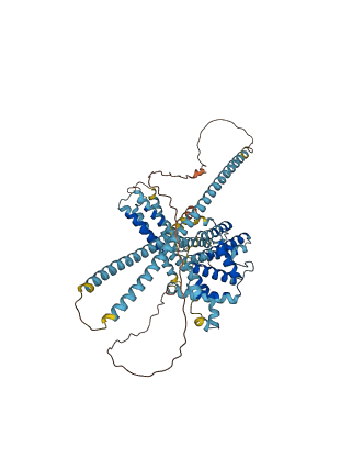
The autoinhibited protein was predicted that may have potential autoinhibitory elements via cis-regPred.
Autoinhibitory domains (AIDs)
Target domain |
|
Relief mechanism |
|
Assay |
cis-regPred |
Accessory elements
No accessory elements
Autoinhibited structure

Activated structure
1 structures for O42869
| Entry ID | Method | Resolution | Chain | Position | Source |
|---|---|---|---|---|---|
| AF-O42869-F1 | Predicted | AlphaFoldDB |
9 variants for O42869
| Variant ID(s) | Position | Change | Description | Diseaes Association | Provenance |
|---|---|---|---|---|---|
| I_3175658_C_T | 90 | A>T | No | Jeffares_SNPs | |
| I_3175508_C_T | 140 | A>T | No | Jeffares_SNPs | |
| I_3175418_T_C | 170 | T>A | No | Jeffares_SNPs | |
| I_3175090_G_A | 279 | A>V | No | Jeffares_SNPs | |
| I_3175067_T_C | 287 | I>V | No | Jeffares_SNPs | |
| I_3174882_T_G | 348 | E>D | No | Jeffares_SNPs | |
| I_3174622_C_T | 435 | R>Q | No | Jeffares_SNPs | |
| I_3174061_G_A | 622 | S>F | No | Jeffares_SNPs | |
| I_3174026_C_T | 634 | A>T | No | Jeffares_SNPs |
No associated diseases with O42869
2 regional properties for O42869
| Type | Name | Position | InterPro Accession |
|---|---|---|---|
| domain | GIT, Spa2 homology (SHD) domain | 33 - 63 | IPR013724-1 |
| domain | GIT, Spa2 homology (SHD) domain | 82 - 112 | IPR013724-2 |
7 GO annotations of cellular component
| Name | Definition |
|---|---|
| actomyosin contractile ring | A cytoskeletal structure composed of actin filaments and myosin that forms beneath the plasma membrane of many cells, including animal cells and yeast cells, in a plane perpendicular to the axis of the spindle, i.e. the cell division plane. In animal cells, the contractile ring is located at the cleavage furrow. In budding fungal cells, e.g. mitotic S. cerevisiae cells, the contractile ring forms at the mother-bud neck before mitosis. |
| cell cortex of growing cell tip | Any cell cortex that is part of a growing cell tip. |
| cell division site | The eventual plane of cell division (also known as cell cleavage or cytokinesis) in a dividing cell. In Eukaryotes, the cleavage apparatus, composed of septin structures and the actomyosin contractile ring, forms along this plane, and the mitotic, or meiotic, spindle is aligned perpendicular to the division plane. In bacteria, the cell division site is generally located at mid-cell and is the site at which the cytoskeletal structure, the Z-ring, assembles. |
| cell tip | The region at the end of the longest axis of a cylindrical or elongated cell. |
| division septum | A cell septum which forms as part of the division site and functions in the compartmentalization of a cell into two daughter cells at division. A division septum spans a cell and does not allow exchange of organelles or cytoplasm between compartments. |
| mitotic actomyosin contractile ring | A cytoskeletal structure composed of actin filaments, myosin, and myosin-associated proteins that forms beneath the plasma membrane of many cells, including animal cells and yeast cells, in a plane perpendicular to the axis of the mitotic spindle, i.e. the cell division plane. Ring contraction is associated with centripetal growth of the membrane that divides the cytoplasm of the two future daughter cells. In animal cells, the mitotic contractile ring is located inside the plasma membrane at the location of the cleavage furrow. In budding fungal cells, e.g. mitotic S. cerevisiae cells, the mitotic contractile ring forms beneath the plasma membrane at the mother-bud neck before mitosis. |
| mitotic actomyosin contractile ring, intermediate layer | The region of the mitotic actomyosin ring in between the proximal layer and the actin filament layer. This region contains the accessory protein network, some actin filaments and connections between the proximal layer and the actin filament layer. |
1 GO annotations of molecular function
| Name | Definition |
|---|---|
| MAP-kinase scaffold activity | The binding activity of a molecule that functions as a physical support for the assembly of a multiprotein mitogen-activated protein kinase (MAPK) complex. Binds multiple kinases of the MAPKKK cascade, and also upstream signaling proteins, permitting those molecules to function in a coordinated way. Bringing together multiple enzymes and their substrates enables the signal to be transduced quickly and efficiently. |
3 GO annotations of biological process
| Name | Definition |
|---|---|
| actin cytoskeleton organization | A process that is carried out at the cellular level which results in the assembly, arrangement of constituent parts, or disassembly of cytoskeletal structures comprising actin filaments and their associated proteins. |
| establishment of cell polarity | The specification and formation of anisotropic intracellular organization or cell growth patterns. |
| establishment or maintenance of cell polarity regulating cell shape | Any cellular process that results in the specification, formation or maintenance of a polarized intracellular organization or cell growth patterns that regulate the shape of a cell. |
No homologous proteins in AiPD
| UniProt AC | Gene Name | Protein Name | Species | Evidence Code |
|---|---|---|---|---|
| No homologous proteins | ||||
| 10 | 20 | 30 | 40 | 50 | 60 |
| MQSIIIRQYR | FLARYLEPEF | VKDPALRNAN | ARPREKLQRL | SHIQFSELLT | DVADELQRRI |
| 70 | 80 | 90 | 100 | 110 | 120 |
| NNDPKVRFLP | PVDSYHPKRN | HARQKLSSLA | PTRLRDLCMD | VFFEVQSRYS | GALKEVSSNT |
| 130 | 140 | 150 | 160 | 170 | 180 |
| PPNMTRAASQ | PPPQVQRLPA | SAPKHDLYSS | ANASSASLHP | TEQRTTSSPT | IPSYANRTHT |
| 190 | 200 | 210 | 220 | 230 | 240 |
| DSPTSLSHRL | PNVPDTNALN | TVQANNSSTS | SLSQGAQTGI | DSSSNYLSRI | EMLESSLAKS |
| 250 | 260 | 270 | 280 | 290 | 300 |
| NSLLDSSKSE | MEALKAKSIS | DATKHKNEIF | QLEEKLHEAS | HEAEISIKKL | NDAENRIKEL |
| 310 | 320 | 330 | 340 | 350 | 360 |
| ENNPTLSFNP | ELEKNLKLMQ | ELIVAESSKT | QHIVELESCI | DSLKAETERW | KKVAINAKSA |
| 370 | 380 | 390 | 400 | 410 | 420 |
| KIDEDIRLFT | MTTVPMKSIR | ELISPNGFLD | EQLYIRYFVE | MNVFVASLRR | DSPSQWMSTA |
| 430 | 440 | 450 | 460 | 470 | 480 |
| KDIALTLEAI | VSSLREKSLL | EELPDLGKKV | DDLCSFTNSL | MEQVRLAVIN | GALLPIYHVD |
| 490 | 500 | 510 | 520 | 530 | 540 |
| ASACSISISL | MDIAKTYGLT | NTGKSSLSVG | RNSLHPVDDL | KALSVAKTLL | SEKSKQFSET |
| 550 | 560 | 570 | 580 | 590 | 600 |
| NQKLLDAISS | DSSSSTIQQR | VHEDINVIRD | TLFDVSSPLE | LMRDRAAYAV | IHNALQEMRR |
| 610 | 620 | 630 | 640 | 650 | |
| YCMQLIEQEG | HGENFGFTDN | NSVPPLTDLA | FSAAKCFKDI | LRAIEDAEFS | VQRTQFSTR |