O42841
Gene name |
SPAC23A1.02c |
Protein name |
Uncharacterized protein C23A1.02c |
Names |
|
Species |
Schizosaccharomyces pombe (strain 972 / ATCC 24843) (Fission yeast) |
KEGG Pathway |
spo:SPAC23A1.02c |
EC number |
|
Protein Class |
|
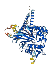
Descriptions
The autoinhibited protein was predicted that may have potential autoinhibitory elements via cis-regPred.
Autoinhibitory domains (AIDs)
Target domain |
|
Relief mechanism |
|
Assay |
cis-regPred |
Accessory elements
No accessory elements
Autoinhibited structure

Activated structure
1 structures for O42841
| Entry ID | Method | Resolution | Chain | Position | Source |
|---|---|---|---|---|---|
| AF-O42841-F1 | Predicted | AlphaFoldDB |
10 variants for O42841
| Variant ID(s) | Position | Change | Description | Diseaes Association | Provenance |
|---|---|---|---|---|---|
| I_4082650_C_G | 42 | K>N | No | Jeffares_SNPs | |
| I_4082360_C_G | 139 | G>A | No | Jeffares_SNPs | |
| I_4082333_A_G | 148 | F>S | No | Jeffares_SNPs | |
| I_4082322_T_C | 152 | N>D | No | Jeffares_SNPs | |
| I_4082184_C_T | 198 | V>I | No | Jeffares_SNPs | |
| I_4082125_G_T,A | 217 | N>K | No | Jeffares_SNPs | |
| I_4082126_T_G | 217 | N>T | No | Jeffares_SNPs | |
| I_4082114_A_G | 221 | I>T | No | Jeffares_SNPs | |
| I_4082102_T_G | 225 | K>T | No | Jeffares_SNPs | |
| I_4081568_G_A | 403 | P>L | No | Jeffares_SNPs |
No associated diseases with O42841
2 GO annotations of cellular component
| Name | Definition |
|---|---|
| endoplasmic reticulum | The irregular network of unit membranes, visible only by electron microscopy, that occurs in the cytoplasm of many eukaryotic cells. The membranes form a complex meshwork of tubular channels, which are often expanded into slitlike cavities called cisternae. The ER takes two forms, rough (or granular), with ribosomes adhering to the outer surface, and smooth (with no ribosomes attached). |
| integral component of endoplasmic reticulum membrane | The component of the endoplasmic reticulum membrane consisting of the gene products and protein complexes having at least some part of their peptide sequence embedded in the hydrophobic region of the membrane. |
1 GO annotations of molecular function
| Name | Definition |
|---|---|
| GPI-mannose ethanolamine phosphate phosphodiesterase activity | A phosphoric diester hydrolase activity that removes the ethanolamine phosphate from mannose 2 of a GPI anchor. |
1 GO annotations of biological process
| Name | Definition |
|---|---|
| GPI anchor biosynthetic process | The chemical reactions and pathways resulting in the formation of a glycosylphosphatidylinositol (GPI) anchor that attaches some membrane proteins to the lipid bilayer of the cell membrane. The phosphatidylinositol group is linked via the C-6 hydroxyl residue of inositol to a carbohydrate chain which is itself linked to the protein via an ethanolamine phosphate group, its amino group forming an amide linkage with the C-terminal carboxyl of the protein. Some GPI anchors have variants on this canonical linkage. |
No homologous proteins in AiPD
| UniProt AC | Gene Name | Protein Name | Species | Evidence Code |
|---|---|---|---|---|
| No homologous proteins | ||||
| 10 | 20 | 30 | 40 | 50 | 60 |
| MFARHPNLLW | LNKQLSILHY | LCLVFLAVYY | AYPLLFGIMP | RKLQLEDENS | FVIMGVADPQ |
| 70 | 80 | 90 | 100 | 110 | 120 |
| IEGNHKIEAN | GFFKGTLDLW | GNDLFLRHLV | HMNQFWGQPD | AMILLGDLVS | FQHLDNEEFN |
| 130 | 140 | 150 | 160 | 170 | 180 |
| KRAKRLKKIT | GAKNFWQVGN | SSLPARTFEN | GNIPVWTIAG | NHDIGYGCES | SDAQISKWEQ |
| 190 | 200 | 210 | 220 | 230 | 240 |
| AMGPVNWVSH | FNVSKFPVRV | IGINSLSLDD | VQFYDANPSD | IINSKSFSSL | GILALSKEAR |
| 250 | 260 | 270 | 280 | 290 | 300 |
| DAWQFLFDIA | LEPSIPTILF | THVPLYKPAN | VCVDEPRIVR | QLDFRVKSQN | HLSYNTTMKI |
| 310 | 320 | 330 | 340 | 350 | 360 |
| FELIPSIKLV | LSGHDHMGCD | YEHPNGAIEH | TLPSAMGYFG | GNIGFVKLIA | TNDVLTESSK |
| 370 | 380 | 390 | 400 | 410 | 420 |
| NTPSVVTFLI | QKLIGQRWKK | ASLKQSKFSS | DIYATYTLSH | GGPSYIWWAL | HISVCVLTIL |
| RLLVISLQHI |