O35405
Gene name |
Pld3 (Sam9) |
Protein name |
5'-3' exonuclease PLD3 |
Names |
Choline phosphatase 3, Phosphatidylcholine-hydrolyzing phospholipase D3, Phospholipase D3, PLD 3, Schwannoma-associated protein 9, SAM-9 |
Species |
Mus musculus (Mouse) |
KEGG Pathway |
mmu:18807 |
EC number |
3.1.16.1: Exonucleases active with either ribo- or deoxyribonucleic acids and producing 3'-phosphomonoesters |
Protein Class |
|
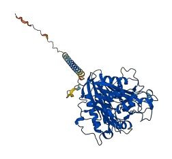
Descriptions
The autoinhibited protein was predicted that may have potential autoinhibitory elements via cis-regPred.
Autoinhibitory domains (AIDs)
Target domain |
|
Relief mechanism |
|
Assay |
cis-regPred |
Accessory elements
No accessory elements
Autoinhibited structure

Activated structure
1 structures for O35405
| Entry ID | Method | Resolution | Chain | Position | Source |
|---|---|---|---|---|---|
| AF-O35405-F1 | Predicted | AlphaFoldDB |
26 variants for O35405
| Variant ID(s) | Position | Change | Description | Diseaes Association | Provenance |
|---|---|---|---|---|---|
| rs3388872349 | 30 | K>M | No | EVA | |
| rs3397860537 | 51 | G>S | No | EVA | |
| rs3397627196 | 52 | A>T | No | EVA | |
| rs3397762498 | 53 | L>M | No | EVA | |
| rs3397716351 | 53 | L>R | No | EVA | |
| rs3397667029 | 54 | M>R | No | EVA | |
| rs3388884778 | 77 | C>Y | No | EVA | |
| rs3388895112 | 78 | Y>F | No | EVA | |
| rs3388884068 | 105 | T>S | No | EVA | |
| rs3388887516 | 108 | A>G | No | EVA | |
| rs3388880932 | 115 | G>D | No | EVA | |
| rs3388892806 | 125 | F>Y | No | EVA | |
| rs3388897260 | 133 | D>E | No | EVA | |
| rs3388865822 | 144 | G>D | No | EVA | |
| rs3388891550 | 168 | P>L | No | EVA | |
| rs3388851341 | 215 | A>G | No | EVA | |
| rs3388884801 | 235 | C>G | No | EVA | |
| rs3388874995 | 238 | L>H | No | EVA | |
| rs3388884069 | 240 | R>P | No | EVA | |
| rs3412895864 | 270 | R>L | No | EVA | |
| rs3388893824 | 292 | S>I | No | EVA | |
| rs3388893860 | 319 | F>L | No | EVA | |
| rs3388865854 | 343 | I>V | No | EVA | |
| rs3388884064 | 367 | H>Q | No | EVA | |
| rs3388884002 | 391 | I>T | No | EVA | |
| rs257620889 | 400 | T>A | No | EVA |
No associated diseases with O35405
3 regional properties for O35405
| Type | Name | Position | InterPro Accession |
|---|---|---|---|
| domain | Phospholipase D/Transphosphatidylase | 194 - 221 | IPR001736-1 |
| domain | Phospholipase D/Transphosphatidylase | 409 - 435 | IPR001736-2 |
| domain | PLD-like domain | 221 - 400 | IPR032803 |
Functions
| Description | ||
|---|---|---|
| EC Number | 3.1.16.1 | Exonucleases active with either ribo- or deoxyribonucleic acids and producing 3'-phosphomonoesters |
| Subcellular Localization |
|
|
| PANTHER Family | ||
| PANTHER Subfamily | ||
| PANTHER Protein Class | ||
| PANTHER Pathway Category | No pathway information available | |
7 GO annotations of cellular component
| Name | Definition |
|---|---|
| early endosome membrane | The lipid bilayer surrounding an early endosome. |
| endoplasmic reticulum membrane | The lipid bilayer surrounding the endoplasmic reticulum. |
| Golgi membrane | The lipid bilayer surrounding any of the compartments of the Golgi apparatus. |
| integral component of membrane | The component of a membrane consisting of the gene products and protein complexes having at least some part of their peptide sequence embedded in the hydrophobic region of the membrane. |
| late endosome membrane | The lipid bilayer surrounding a late endosome. |
| lysosomal lumen | The volume enclosed within the lysosomal membrane. |
| lysosomal membrane | The lipid bilayer surrounding the lysosome and separating its contents from the cell cytoplasm. |
2 GO annotations of molecular function
| Name | Definition |
|---|---|
| phospholipase D activity | Catalysis of the reaction: a phosphatidylcholine + H2O = choline + a phosphatidate. |
| single-stranded DNA 5'-3' exodeoxyribonuclease activity | Catalysis of the sequential cleavage of nucleotides (such as mononucleotides or dinucleotides) from a free 5' terminus of a single-stranded DNA molecule. |
4 GO annotations of biological process
| Name | Definition |
|---|---|
| inflammatory response | The immediate defensive reaction (by vertebrate tissue) to infection or injury caused by chemical or physical agents. The process is characterized by local vasodilation, extravasation of plasma into intercellular spaces and accumulation of white blood cells and macrophages. |
| innate immune response | Innate immune responses are defense responses mediated by germline encoded components that directly recognize components of potential pathogens. |
| myotube differentiation | The process in which a relatively unspecialized cell acquires specialized features of a myotube cell. Myotube differentiation starts with myoblast fusion and the appearance of specific cell markers (this is the cell development step). Then individual myotubes can fuse to form bigger myotubes and start to contract. Myotubes are multinucleated cells that are formed when proliferating myoblasts exit the cell cycle, differentiate and fuse. |
| regulation of cytokine production involved in inflammatory response | Any process that modulates the frequency, rate or extent of cytokine production involved in inflammatory response. |
No homologous proteins in AiPD
| UniProt AC | Gene Name | Protein Name | Species | Evidence Code |
|---|---|---|---|---|
| No homologous proteins | ||||
| 10 | 20 | 30 | 40 | 50 | 60 |
| MKPKLMYQEL | KVPVEEPAGE | LPLNEIEAWK | AAEKKARWVL | LVLILAVVGF | GALMTQLFLW |
| 70 | 80 | 90 | 100 | 110 | 120 |
| EYGDLHLFGP | NQRPAPCYDP | CEAVLVESIP | EGLEFPNATT | SNPSTSQAWL | GLLAGAHSSL |
| 130 | 140 | 150 | 160 | 170 | 180 |
| DIASFYWTLT | NNDTHTQEPS | AQQGEEVLQQ | LQALAPRGVK | VRIAVSKPNG | PLADLQSLLQ |
| 190 | 200 | 210 | 220 | 230 | 240 |
| SGAQVRMVDM | QKLTHGVLHT | KFWVVDQTHF | YLGSANMDWR | SLTQVKELGV | VMYNCSCLAR |
| 250 | 260 | 270 | 280 | 290 | 300 |
| DLTKIFEAYW | FLGQAGSSIP | STWPRSFDTR | YNQETPMEIC | LNGTPALAYL | ASAPPPLCPS |
| 310 | 320 | 330 | 340 | 350 | 360 |
| GRTPDLKALL | NVVDSARSFI | YIAVMNYLPT | MEFSHPRRFW | PAIDDGLRRA | AYERGVKVRL |
| 370 | 380 | 390 | 400 | 410 | 420 |
| LISCWGHSDP | SMRSFLLSLA | ALHDNHTHSD | IQVKLFVVPT | DESQARIPYA | RVNHNKYMVT |
| 430 | 440 | 450 | 460 | 470 | 480 |
| ERASYIGTSN | WSGSYFTETA | GTSLLVTQNG | HGGLRSQLEA | VFLRDWESPY | SHDLDTSANS |
| VGNACRLL |